Bone Scan Timing: When to Acquire Delayed Images

Public domain · Wikimedia Commons · source file
Question
When performing a Tc-99m MDP whole-body bone scan, images are typically acquired how long after intravenous injection?
- AImmediately after injection
- B30 minutes after injection
- C6–8 hours after injection
- D2–4 hours after injection
Show the answer and explanation
D. 2–4 hours after injection
Whole-body bone scan 'delayed phase' images are typically acquired 2–4 hours after IV injection of Tc-99m MDP. This delay allows: (1) adequate time for bone uptake via chemisorption onto hydroxyapatite; (2) sufficient clearance of background soft-tissue and blood-pool activity via renal excretion; (3) optimal bone-to-soft tissue contrast. Imaging too early results in high background soft-tissue activity and poor bone-to-background ratio. In patients with renal impairment, the delay may need to be extended to 4–6 hours to allow adequate clearance.
More bone imaging images
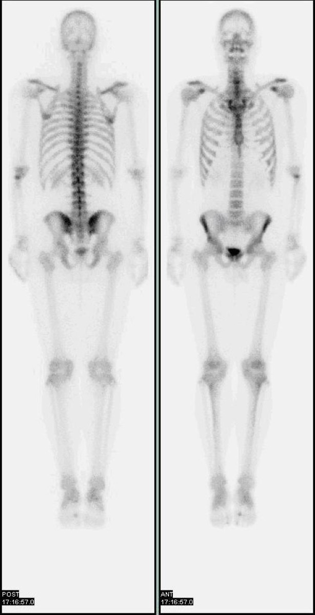 Normal Whole-Body Bone Scan (Tc-99m MDP)
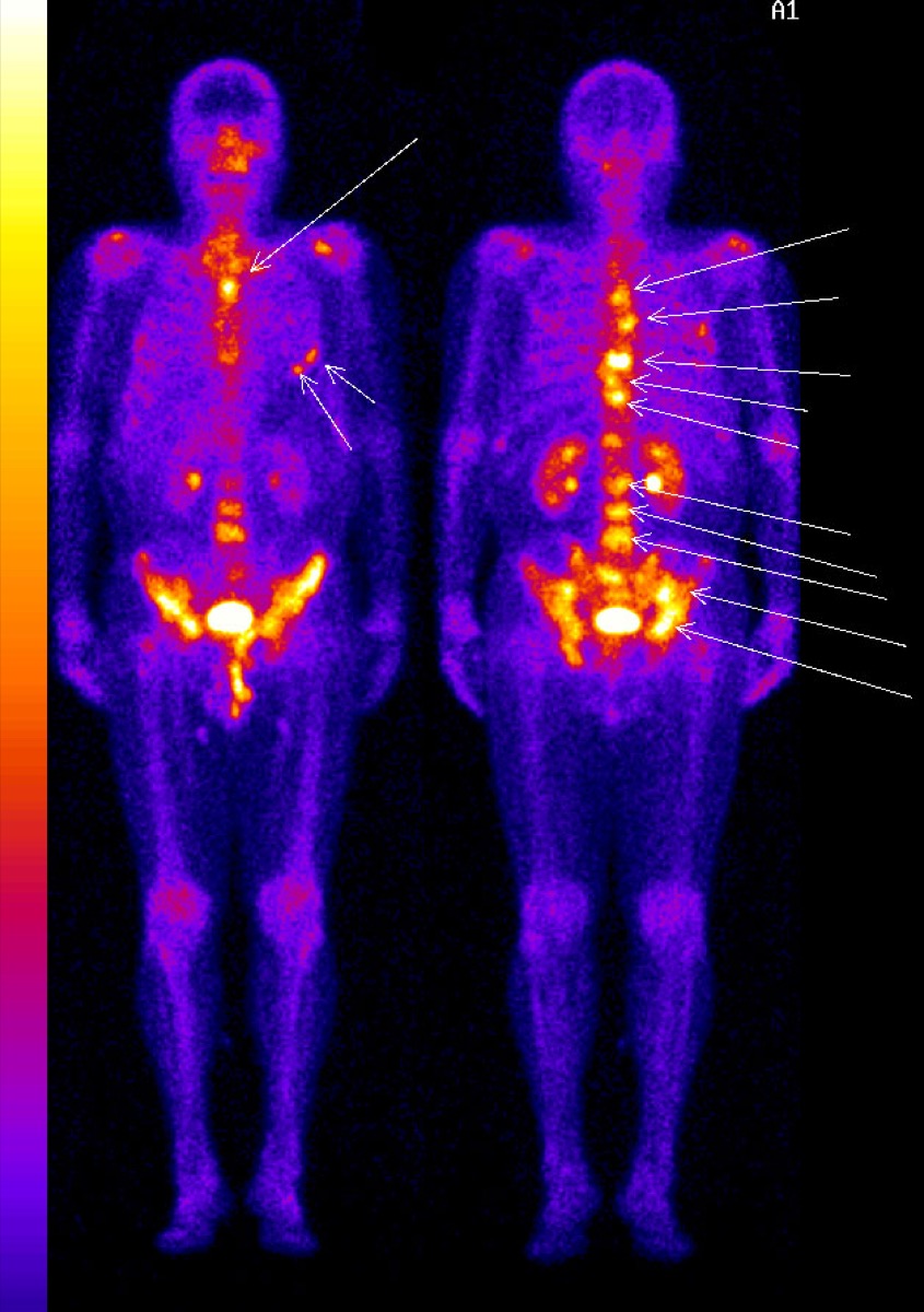 Diffuse Skeletal Metastases on Bone Scan
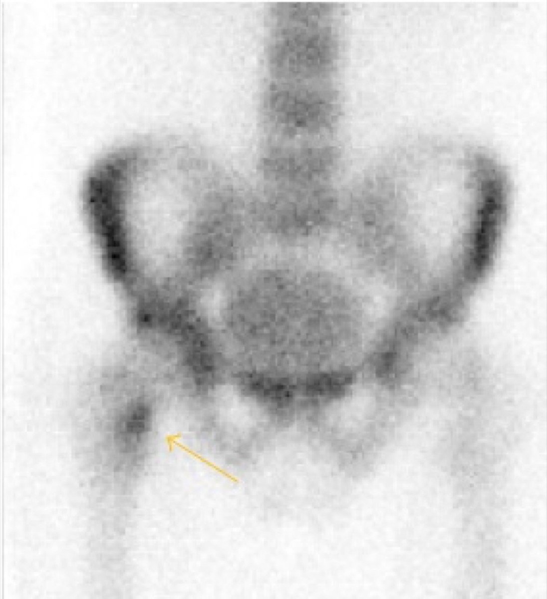 Femoral Neck Stress Fracture on Bone Scan
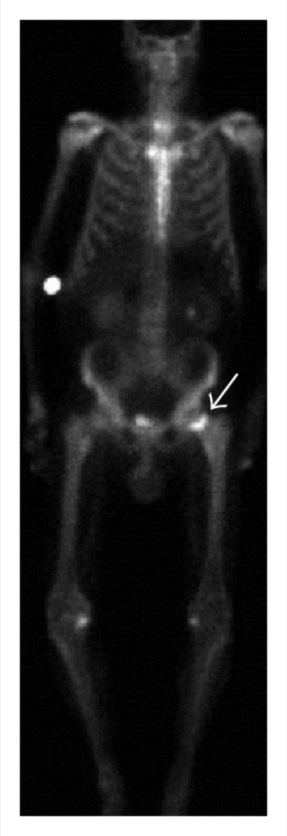 Femoral Neck Insufficiency Fracture on Bone Scan
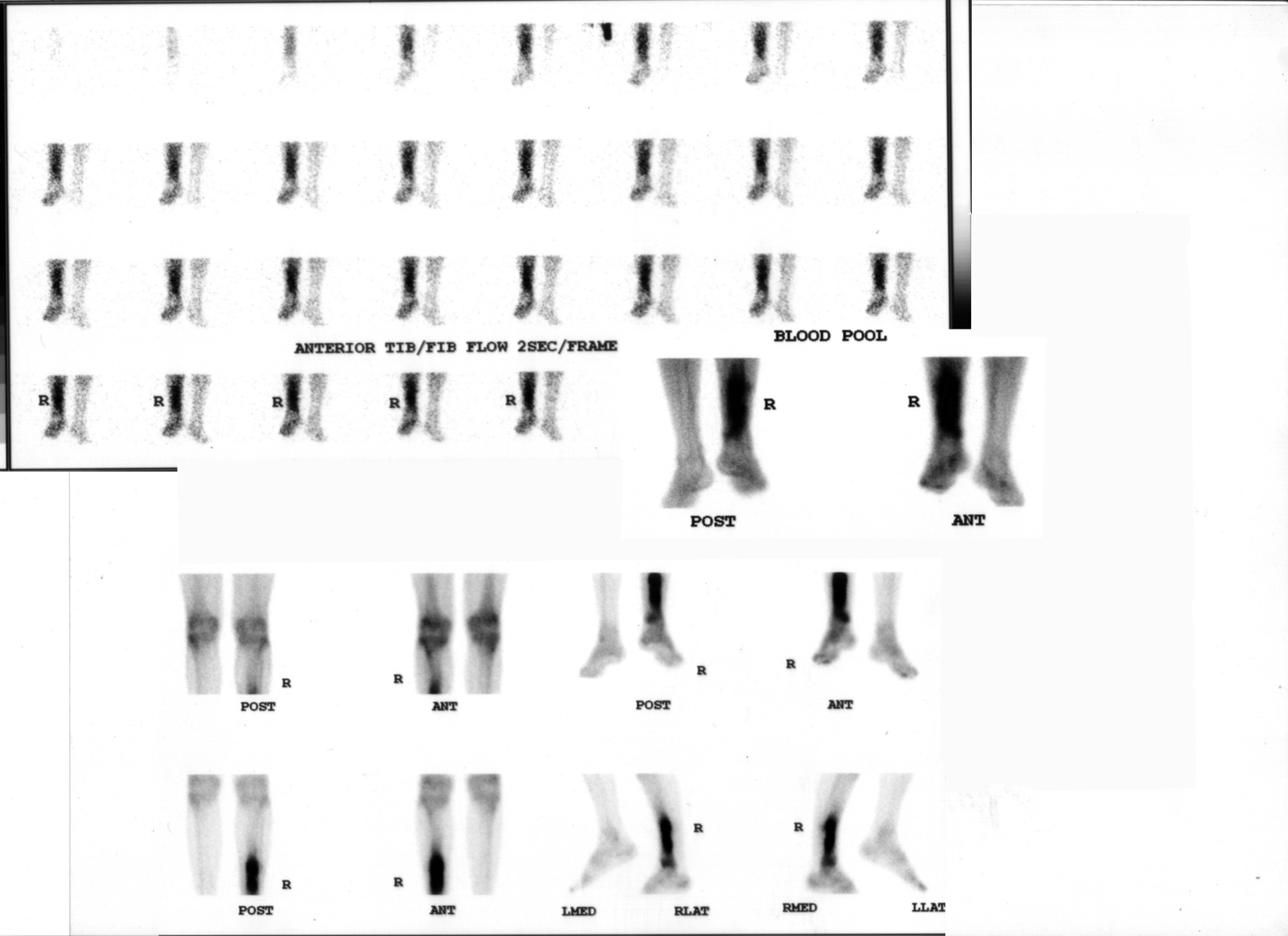 Osteomyelitis on Three-Phase Bone Scan
How Tc-99m MDP Localizes in Bone
All 130 nuclear medicine images by category →
Practise image interpretation in the app
All 130 images as an interactive quiz, alongside 500 NMTCB and ARRT(N) questions with worked explanations, case studies and timed mock exams. 25 questions are free, no card and no account.