Femoral Neck Stress Fracture on Bone Scan
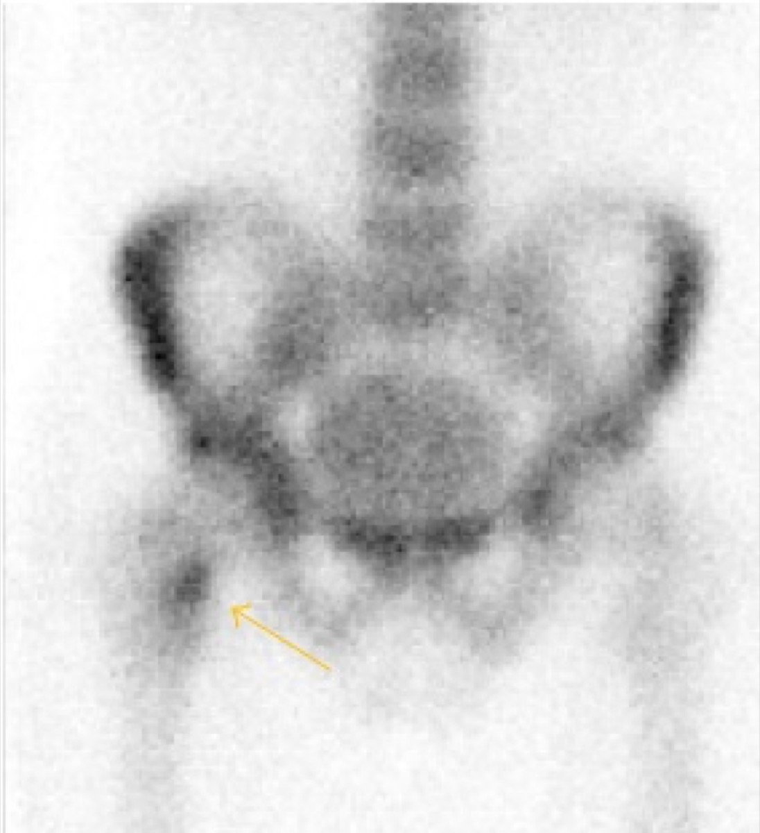
CC BY 4.0 · Santiago et al. / Wikimedia Commons · source file
Question
This bone scan shows a linear focus of intense increased uptake in the proximal femur/femoral neck region in an athlete presenting with hip pain. No abnormality was visible on initial plain radiographs. This pattern is most consistent with:
- AStress fracture
- BAvascular necrosis of the femoral head
- CMetastatic lesion
- DTransient osteoporosis
Show the answer and explanation
A. Stress fracture
Bone scintigraphy is highly sensitive for detecting stress fractures that are radiographically occult (invisible on plain X-ray). A stress fracture typically appears as a linear or fusiform focus of intense increased uptake at the cortex. The clinical context of an athlete with hip pain and a negative radiograph makes stress fracture the most likely diagnosis. Avascular necrosis would typically show a cold (photopenic) area early. A metastasis would be in the medullary space and not linear.
More bone imaging images
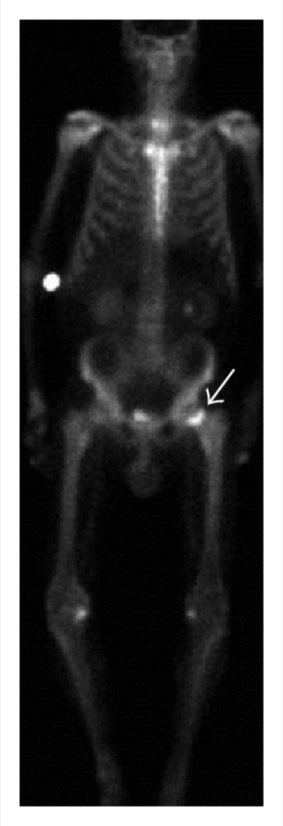 Femoral Neck Insufficiency Fracture on Bone Scan
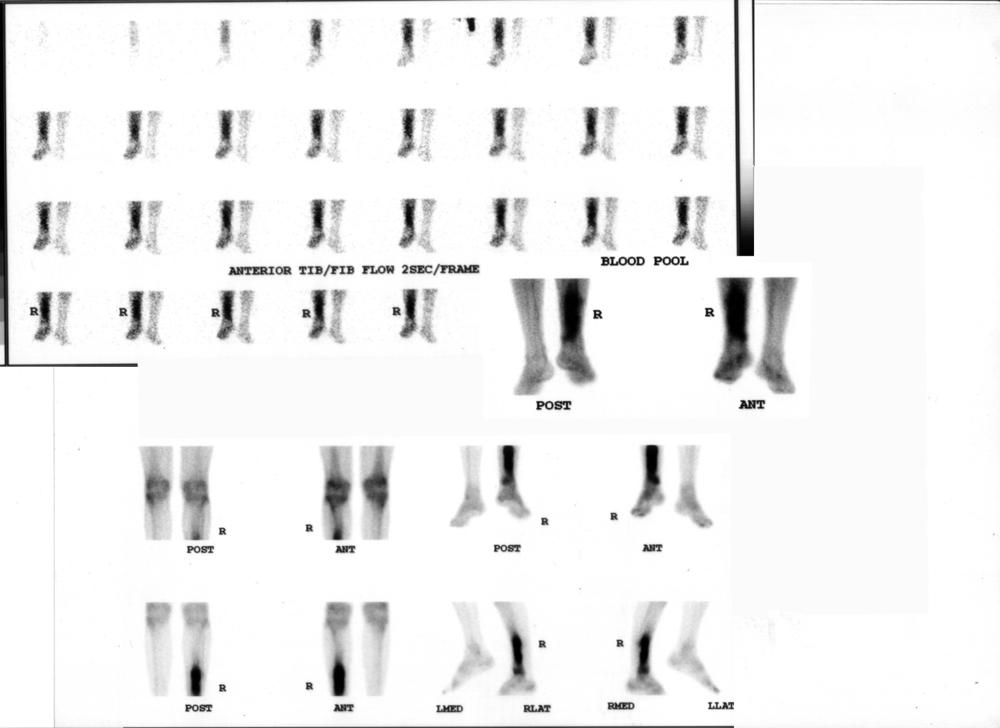 Osteomyelitis on Three-Phase Bone Scan
 How Tc-99m MDP Localizes in Bone
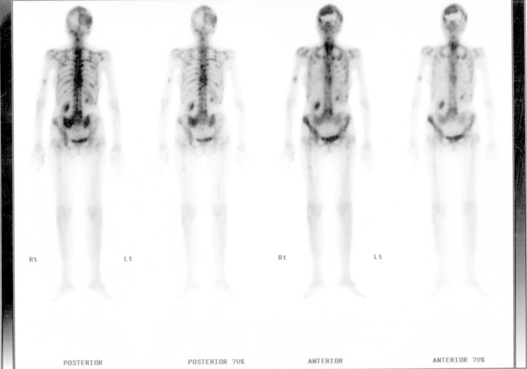 Bone Metastases: Prostate and Breast Primaries
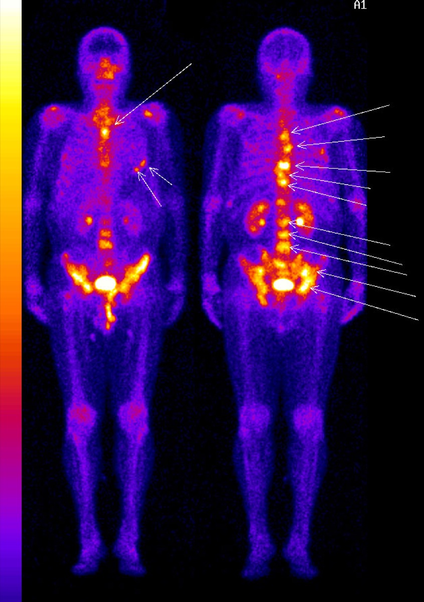 Superscan on Bone Scintigraphy
Paget’s Disease of Bone on Bone Scan
All 130 nuclear medicine images by category →
Practise image interpretation in the app
All 130 images as an interactive quiz, alongside 500 NMTCB and ARRT(N) questions with worked explanations, case studies and timed mock exams. 25 questions are free, no card and no account.