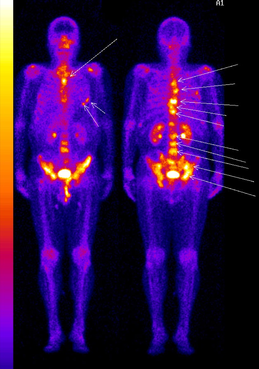
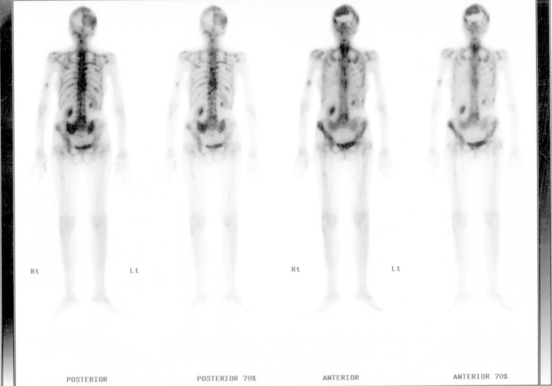
Diffuse Skeletal Metastases on Bone Scan
CC BY-SA 3.0 · Wikimedia Commons · source file
Question
This whole-body bone scan shows multiple foci of abnormally increased tracer uptake scattered throughout the axial skeleton — spine, ribs, pelvis, and skull. In a patient with known carcinoma, this pattern is most consistent with:
- AMultifocal osteomyelitis
- BHyperparathyroidism
- CDiffuse skeletal metastases
- DPolyostotic Paget's disease
Show the answer and explanation
C. Diffuse skeletal metastases
Multiple randomly distributed foci of increased uptake throughout the axial and appendicular skeleton in a patient with known primary cancer is the classic pattern of diffuse skeletal metastases. Prostate and breast cancer most commonly cause osteoblastic (hot) metastases on bone scan. Paget's disease shows intense uptake in expanded bone segments. Hyperparathyroidism may cause a 'superscan' with generalized increased uptake but typically lacks the focal random distribution seen here.
More bone imaging images
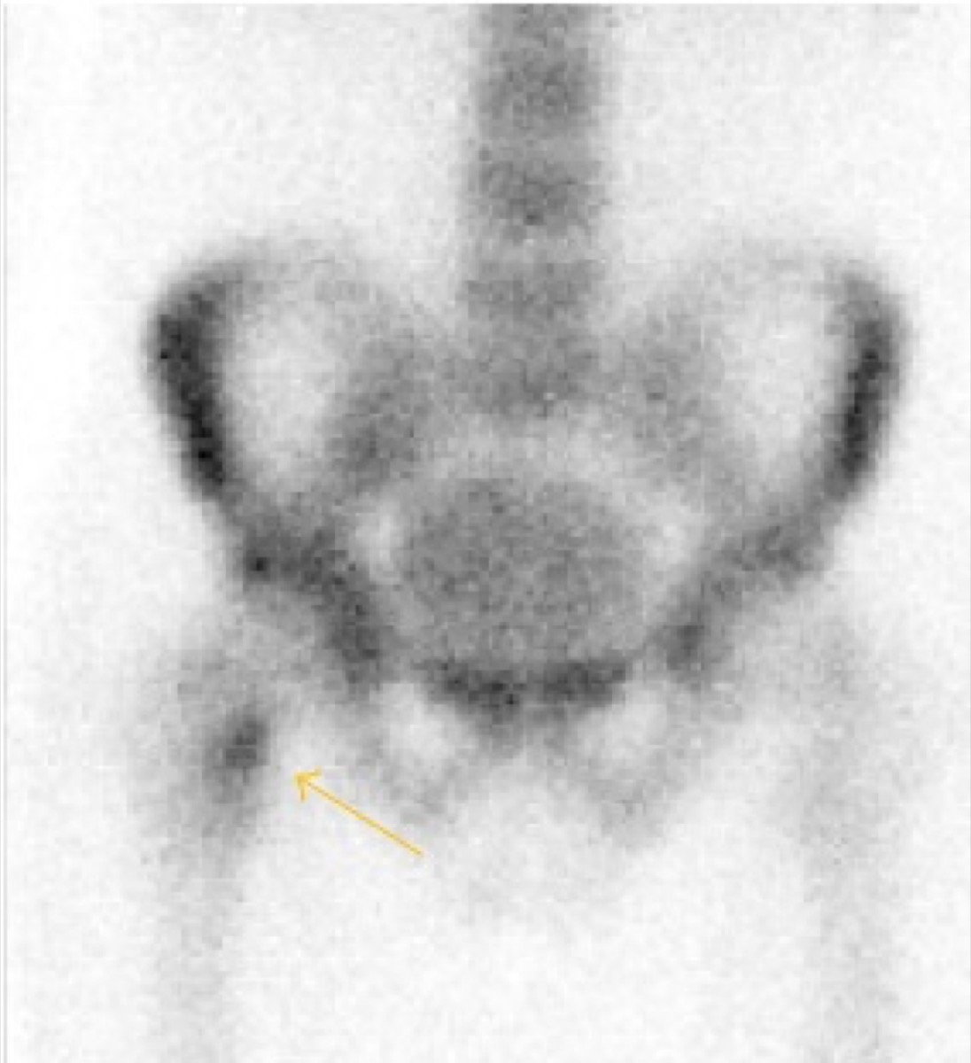 Femoral Neck Stress Fracture on Bone Scan
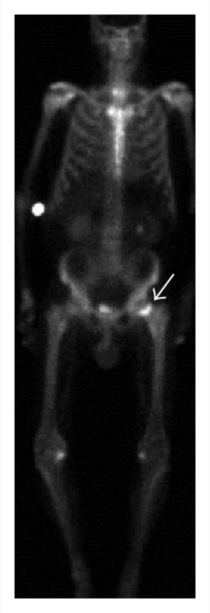 Femoral Neck Insufficiency Fracture on Bone Scan
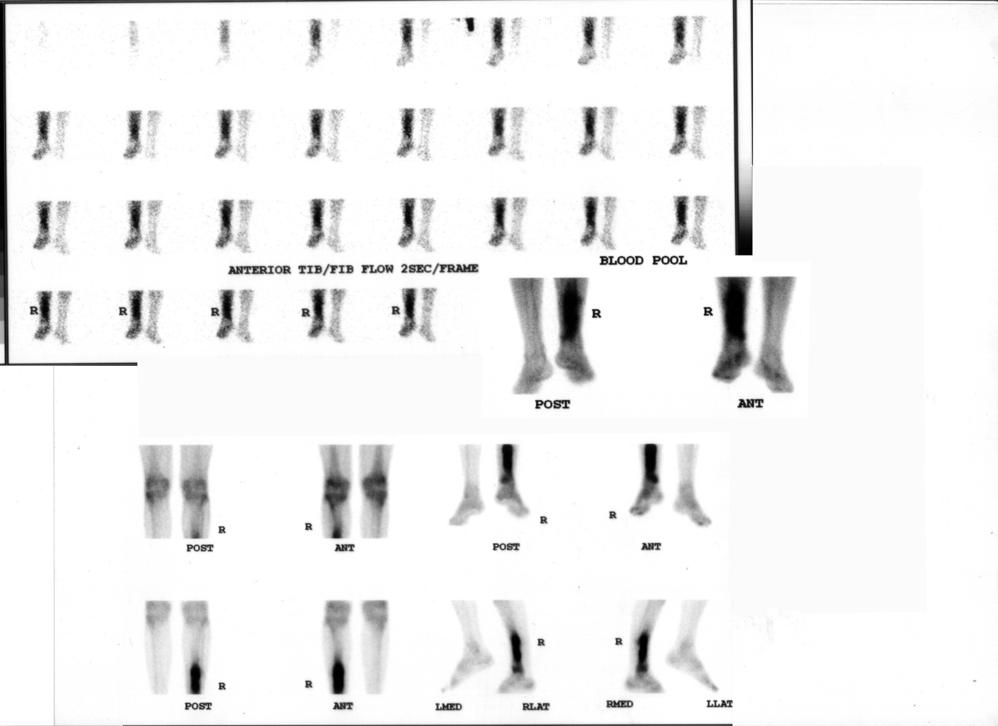 Osteomyelitis on Three-Phase Bone Scan
 How Tc-99m MDP Localizes in Bone
 Bone Metastases: Prostate and Breast Primaries
Superscan on Bone Scintigraphy
All 130 nuclear medicine images by category →
Practise image interpretation in the app
All 130 images as an interactive quiz, alongside 500 NMTCB and ARRT(N) questions with worked explanations, case studies and timed mock exams. 25 questions are free, no card and no account.