CT Attenuation Correction Misregistration in Cardiac SPECT
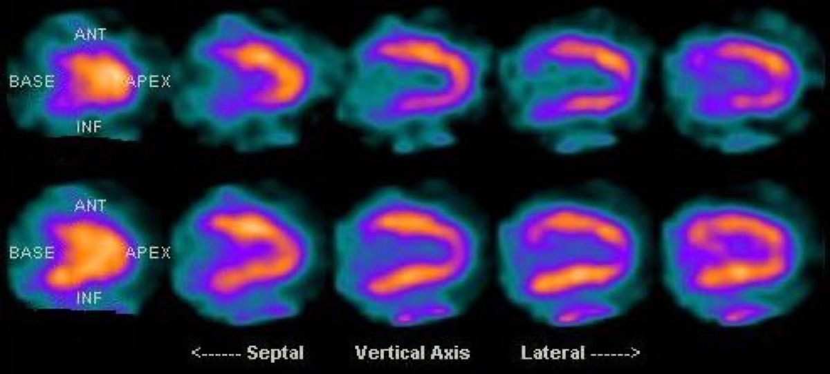
Public domain · KieranMaher / Wikimedia Commons · source file
Question
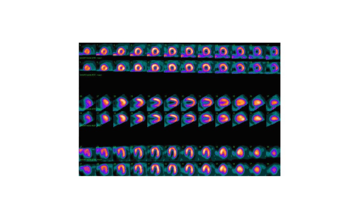
CT-based attenuation correction (AC) improves cardiac SPECT image quality by correcting for photon attenuation in overlying soft tissue. However, AC can also introduce artifacts. Which of the following is a recognized PITFALL of CT attenuation correction in cardiac SPECT?
- ACT attenuation correction eliminates all perfusion defects including true ischemia
- BAC-corrected scans always show inferior wall defects
- CCT attenuation correction reduces LVEF calculation accuracy
- DMotion misregistration between CT and SPECT acquisition can create artifactual perfusion defects
Show the answer and explanation
D. Motion misregistration between CT and SPECT acquisition can create artifactual perfusion defects
CT attenuation correction in cardiac SPECT requires spatial alignment (registration) of the CT map with the SPECT data. If the patient moves between the CT and SPECT acquisitions (e.g., from cardiac motion or patient repositioning), the CT map will not perfectly correspond to the SPECT image, creating artifactual perfusion defects — typically in the anterior or inferior wall. This motion misregistration artifact is a recognized pitfall of AC-corrected cardiac SPECT. It is identified by comparing AC-corrected and non-corrected images side by side; a defect present only on AC images but not on non-corrected images may be an AC artifact.
More cardiac imaging images
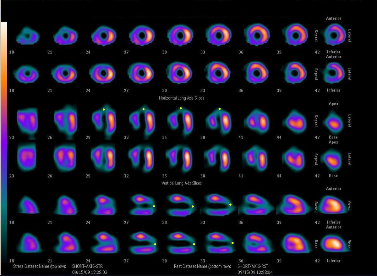 FDG PET Viability: Hibernating Myocardium
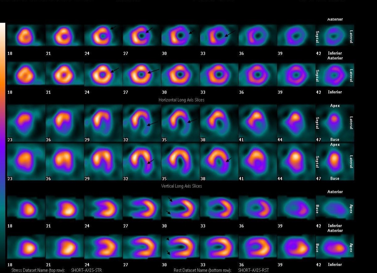 Inferior Wall Ischemia: Right Coronary Artery Territory
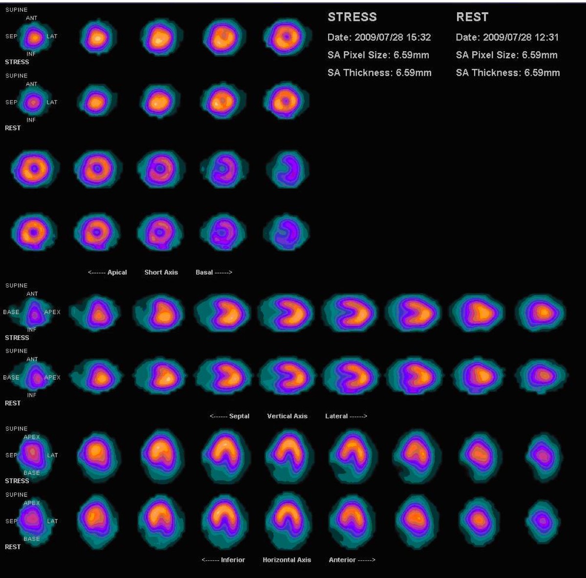 Vasodilator Stress Contraindications: Caffeine and Bronchospasm
 Gated SPECT: Ejection Fraction and Ventricular Volumes
LAD Territory Perfusion Defect on SPECT
Multivessel Coronary Disease on Stress MPI
All 130 nuclear medicine images by category →
Practise image interpretation in the app
All 130 images as an interactive quiz, alongside 500 NMTCB and ARRT(N) questions with worked explanations, case studies and timed mock exams. 25 questions are free, no card and no account.