Diaphragmatic Attenuation Artifact on Cardiac SPECT
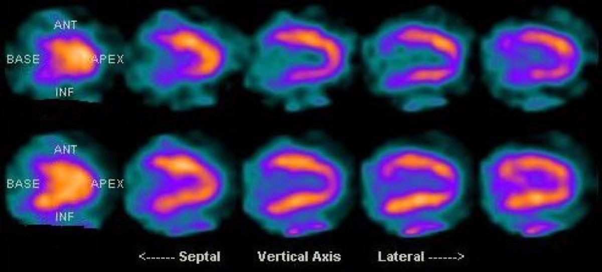
Public domain · KieranMaher / Wikimedia Commons · source file
Question
These cardiac SPECT images show an apparent inferior wall perfusion defect on the non-attenuation-corrected images (top) that is no longer visible on the attenuation-corrected images (bottom). What is the most likely cause of the apparent inferior wall defect on the non-corrected images?
- ADiaphragmatic attenuation artifact
- BRight coronary artery (RCA) territory ischemia
- CPrior inferior wall myocardial infarction
- DPatient motion during acquisition
Show the answer and explanation
A. Diaphragmatic attenuation artifact
Diaphragmatic attenuation is the most common cause of apparent inferior wall defects on non-corrected cardiac SPECT, particularly in male patients. The diaphragm overlies and attenuates photons from the inferior wall of the left ventricle, creating a false-positive perfusion defect. Attenuation correction (using CT or transmission scan data) compensates for this, and the defect disappears — confirming it as artifact rather than true ischemia or infarction.
More cardiac imaging images
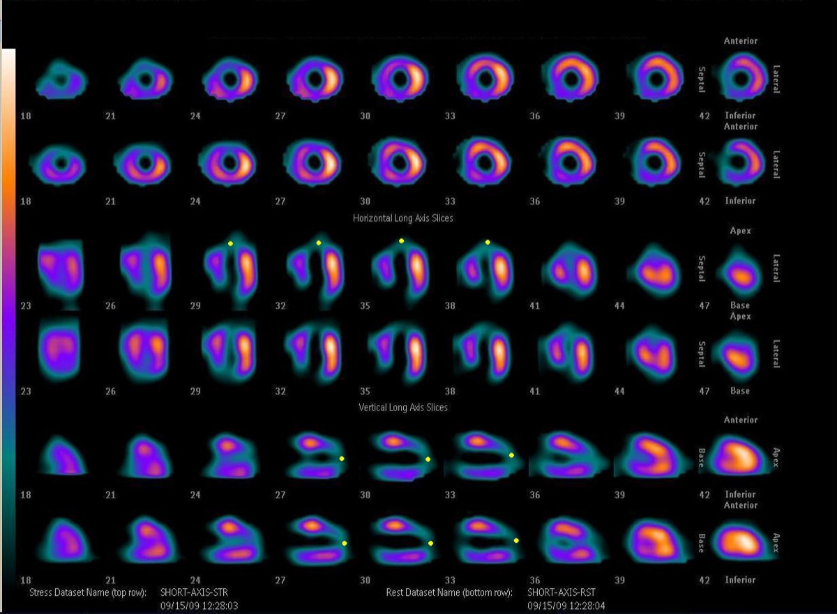 Fixed Perfusion Defect: Myocardial Scar on SPECT
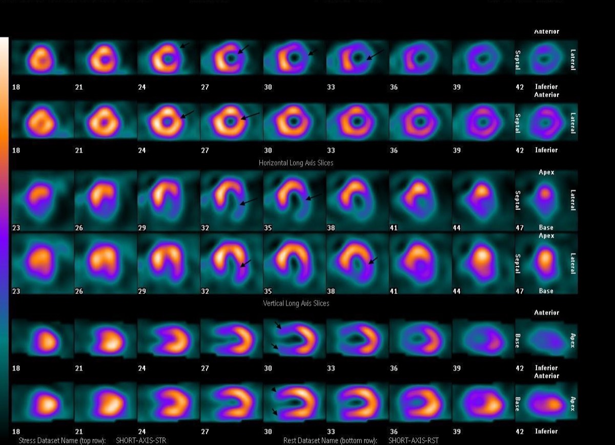 Reversible Lateral Wall Defect: Circumflex Ischemia
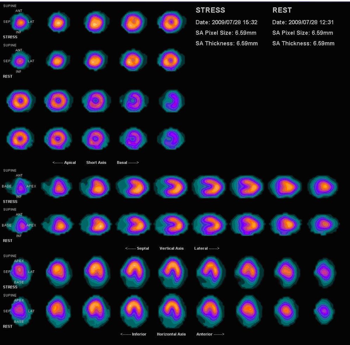 Bull’s-Eye Polar Map: What the Center Shows
Anteroapical Fixed Defect: LAD Infarction on MPI
Transient Ischemic Dilation (TID) on Gated SPECT
Rb-82 PET Perfusion and the Sr-82/Rb-82 Generator
All 130 nuclear medicine images by category →
Practise image interpretation in the app
All 130 images as an interactive quiz, alongside 500 NMTCB and ARRT(N) questions with worked explanations, case studies and timed mock exams. 25 questions are free, no card and no account.