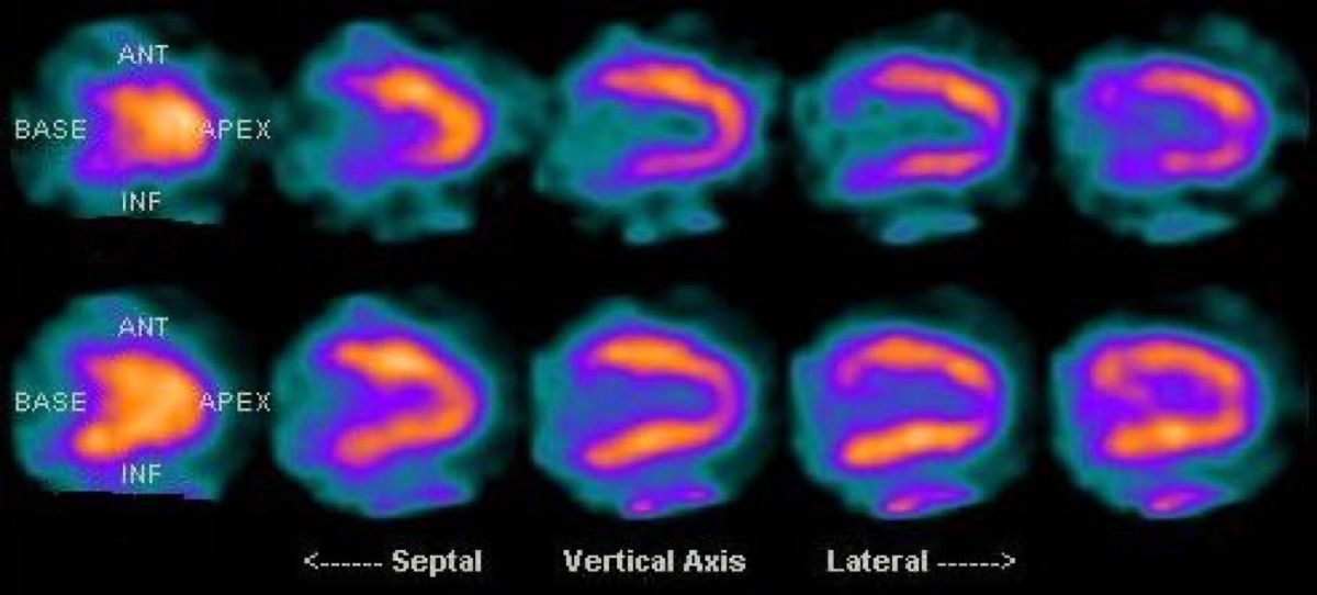
Bull’s-Eye Polar Map: What the Center Shows
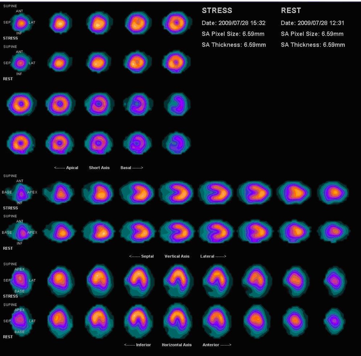
CC BY 3.0 · Myohan / Wikimedia Commons · source file
Question
On a myocardial perfusion SPECT study, the bull's-eye (polar map) display shows the entire left ventricular myocardium in a single image. The CENTER of the polar map corresponds to which myocardial region?
- AThe basal septum
- BThe lateral free wall
- CThe posterior wall
- DThe cardiac apex
Show the answer and explanation
D. The cardiac apex
The bull's-eye (polar map) displays the entire left ventricular myocardium as a flat circular projection: the CENTER represents the APEX, and the outer RING represents the basal segments. Moving outward from center to periphery corresponds to moving from apex to base. The polar map is divided into coronary artery territories: anterior (LAD), inferior (RCA), and lateral (LCX). This display facilitates visual assessment and comparison to normal databases for automated quantification of perfusion defects.
More cardiac imaging images
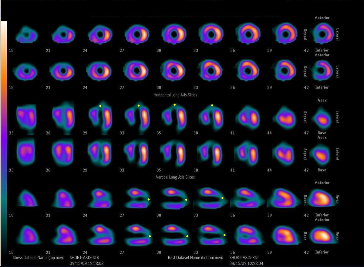 Anteroapical Fixed Defect: LAD Infarction on MPI
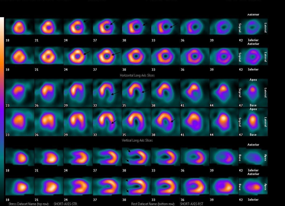 Transient Ischemic Dilation (TID) on Gated SPECT
Rb-82 PET Perfusion and the Sr-82/Rb-82 Generator
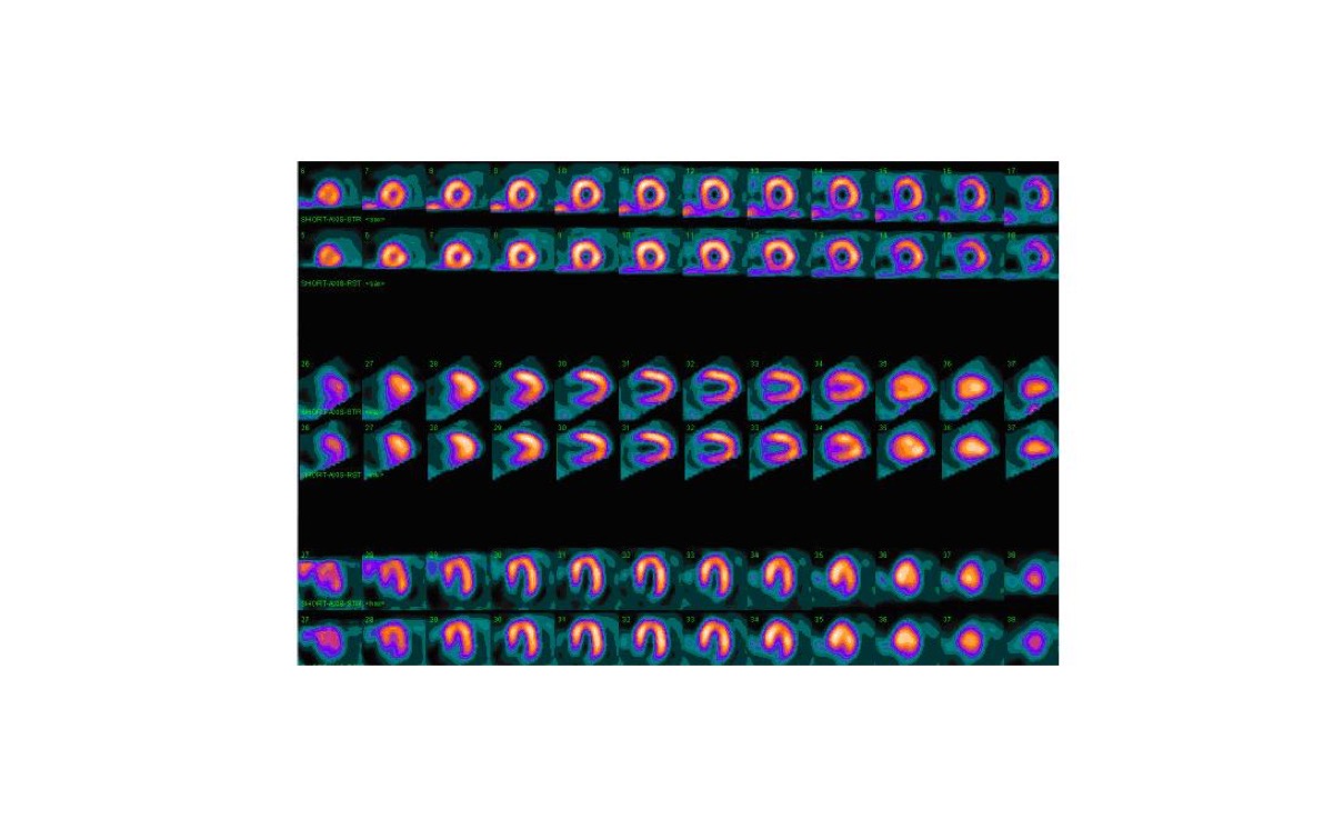 Breast Attenuation Artifact on Myocardial Perfusion SPECT
 CT Attenuation Correction Misregistration in Cardiac SPECT
FDG PET Viability: Hibernating Myocardium
All 130 nuclear medicine images by category →
Practise image interpretation in the app
All 130 images as an interactive quiz, alongside 500 NMTCB and ARRT(N) questions with worked explanations, case studies and timed mock exams. 25 questions are free, no card and no account.