PET Coincidence Detection of 511 keV Photons
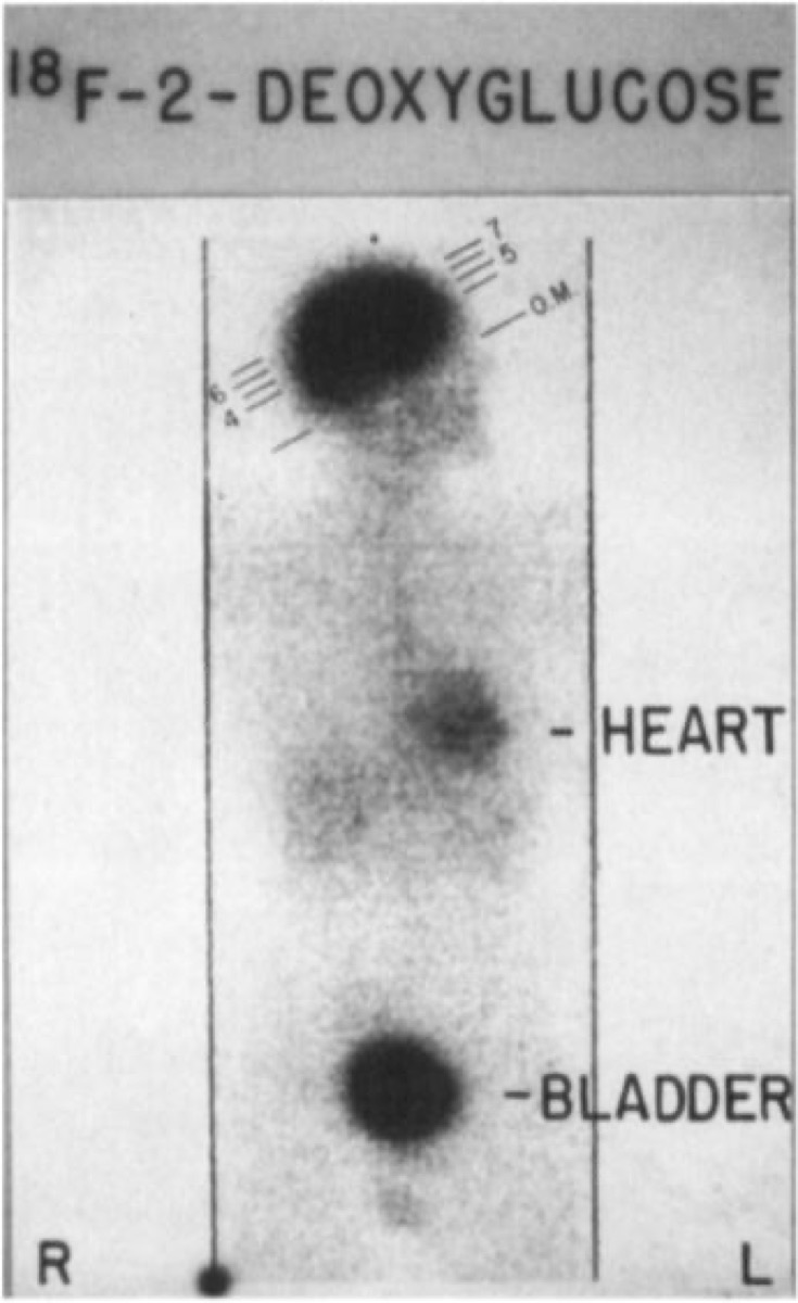
CC BY-SA 4.0 · Pennradiology / Wikimedia Commons · source file
Question
PET imaging detects gamma photons produced by positron annihilation. What energy photons are detected, and how many are produced per annihilation event?
- AOne 140 keV photon per decay event
- BFour 511 keV photons in random directions
- COne 511 keV photon detected by a single crystal
- DTwo 511 keV photons emitted in exactly opposite directions (180°) — detected simultaneously (coincidence detection)
Show the answer and explanation
D. Two 511 keV photons emitted in exactly opposite directions (180°) — detected simultaneously (coincidence detection)
PET imaging is based on positron-electron annihilation physics. When a positron (β+) is emitted and travels a short distance (range), it collides with an electron. This annihilation produces TWO 511 keV gamma photons emitted in EXACTLY OPPOSITE DIRECTIONS (180° apart). PET scanners detect these two photons simultaneously (within a coincidence timing window of ~3–10 ns) using opposing detector rings — a process called coincidence detection. This coincidence detection provides electronic collimation (no physical collimator needed), far superior sensitivity compared to SPECT. The 511 keV photon energy corresponds to the rest-mass energy of an electron (0.511 MeV = m_e c²).
More pet and oncology images
 MIBG Scan for Neuroblastoma Staging
 Lu-177 DOTATATE Therapy for Neuroendocrine Tumors
Paraganglioma: Ga-68 DOTATATE PET vs MIBG
Medications to Stop Before an MIBG Scan
Carcinoid Tumor on Somatostatin Receptor Imaging
 OctreoScan: Why Delayed 24-Hour Images
All 130 nuclear medicine images by category →
Practise image interpretation in the app
All 130 images as an interactive quiz, alongside 500 NMTCB and ARRT(N) questions with worked explanations, case studies and timed mock exams. 25 questions are free, no card and no account.