Osteomyelitis on Three-Phase Bone Scan
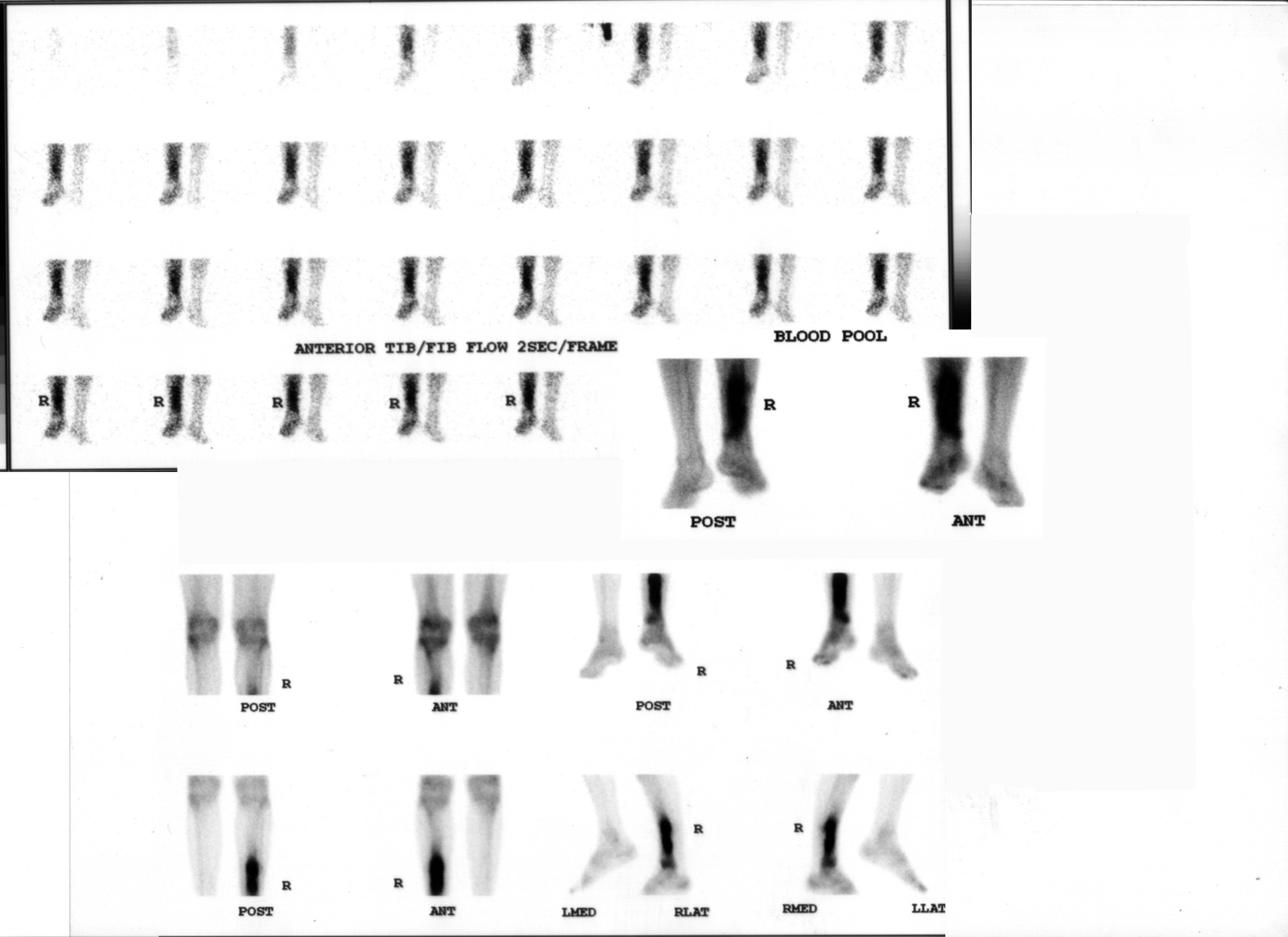
Provided clinical image
Question
A three-phase bone scan shows increased uptake in ALL three phases (flow, blood pool, and delayed) in a painful, swollen extremity following trauma. No radiographic fracture is seen. This pattern is most characteristic of:
- AOsteomyelitis
- BCellulitis
- CComplex Regional Pain Syndrome (CRPS / RSD)
- DDeep vein thrombosis
Show the answer and explanation
A. Osteomyelitis
Osteomyelitis characteristically shows increased uptake in ALL THREE phases of bone scintigraphy: (1) FLOW phase — increased radionuclide angiogram reflecting hyperemia; (2) BLOOD POOL phase — increased activity from vascular pooling in inflamed tissue; (3) DELAYED phase — increased bone uptake from osteoblastic activity and periosteal reaction. This tri-phasic positivity is the hallmark of osteomyelitis. Cellulitis shows increased flow and blood pool phases but NORMAL delayed phase (soft-tissue hyperemia only, no bone involvement). CRPS can show diffuse periarticular uptake on all three phases but in a different distribution and clinical context. DVT does not significantly affect bone scan phases.
More bone imaging images
 How Tc-99m MDP Localizes in Bone
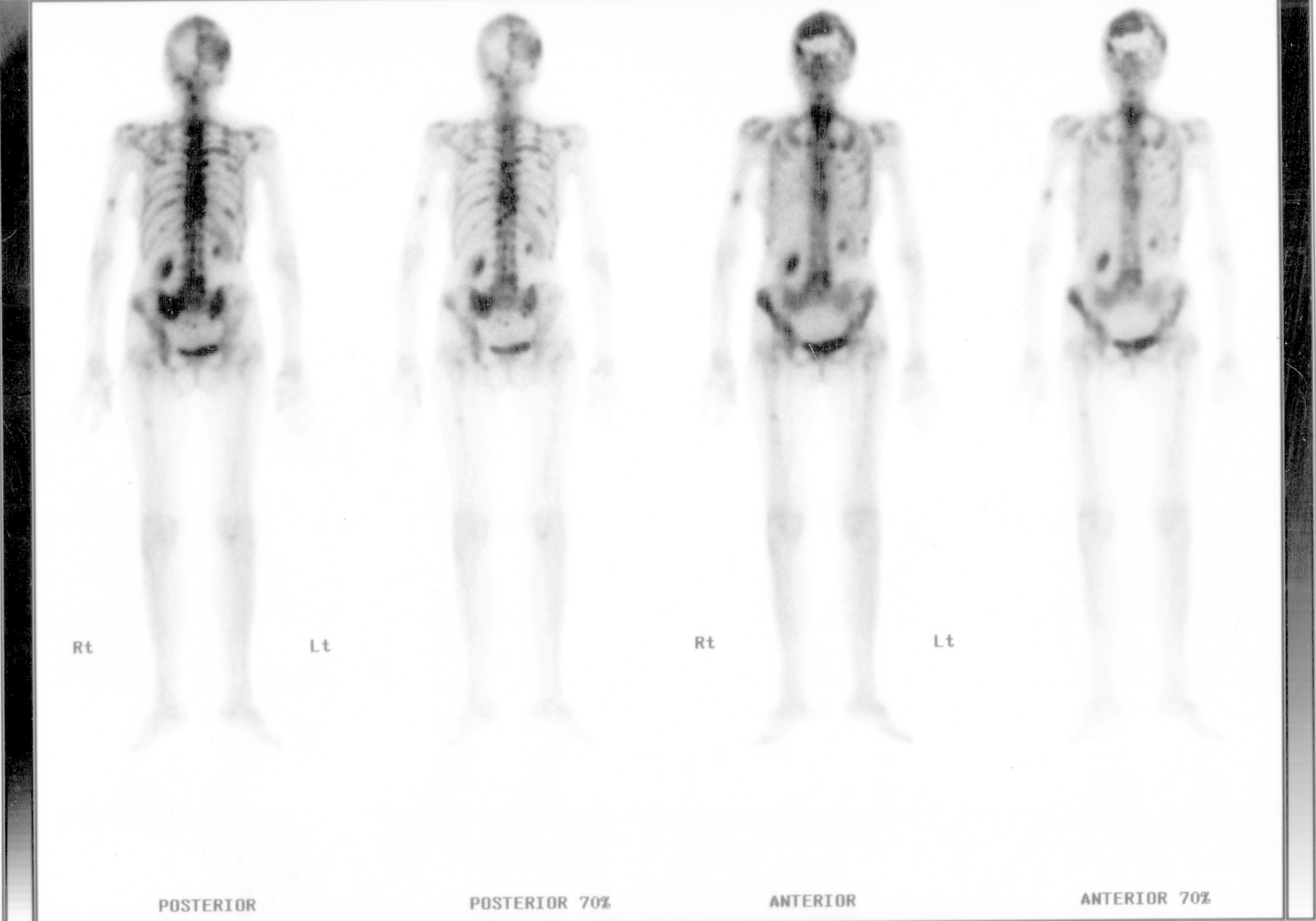 Bone Metastases: Prostate and Breast Primaries
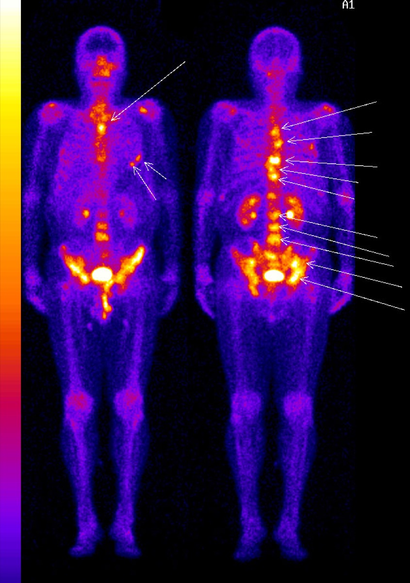 Superscan on Bone Scintigraphy
Paget’s Disease of Bone on Bone Scan
Photopenic Bone Lesions: Multiple Myeloma
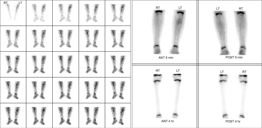 Cellulitis vs Osteomyelitis on Three-Phase Bone Scan
All 130 nuclear medicine images by category →
Practise image interpretation in the app
All 130 images as an interactive quiz, alongside 500 NMTCB and ARRT(N) questions with worked explanations, case studies and timed mock exams. 25 questions are free, no card and no account.