Normal Hepatobiliary Scan Sequence (Mebrofenin)
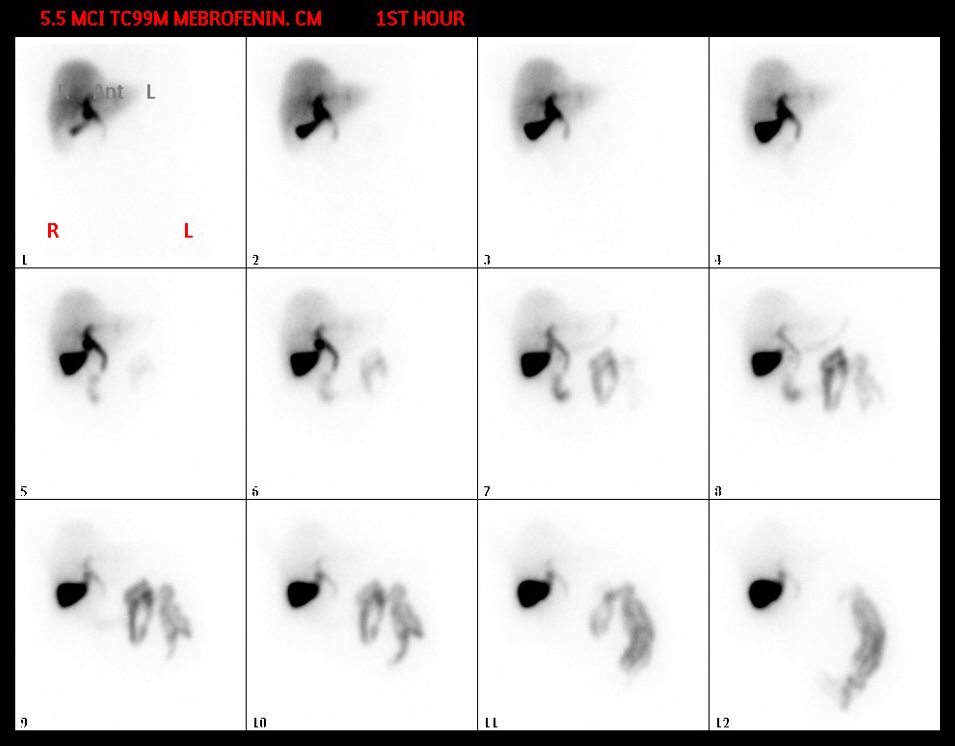
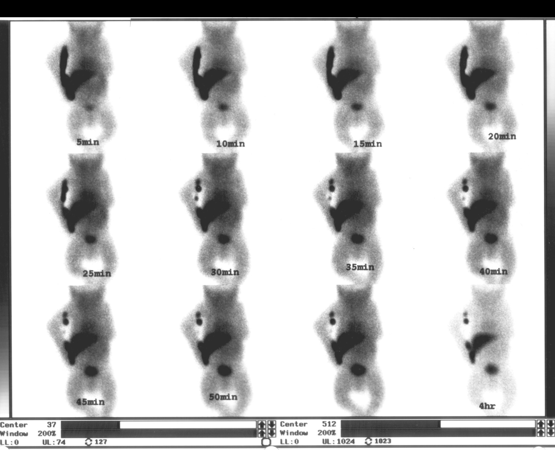
Provided clinical image
Question
This dynamic series shows 12 sequential frames acquired over the first hour after injection of 5.5 mCi Tc-99m mebrofenin. What is the expected normal sequence of organ visualization in a properly functioning study?
- AKidneys → bladder → bowel, with the liver serving only as a transit organ and showing minimal retained activity by the sixth frame
- BLiver uptake first, followed by the bile ducts, then the gallbladder and bowel — either can appear before the other, but both should be visualized within 60 minutes in a normal study
- CHeart and blood pool → liver → delayed small bowel activity that appears only after 3–4 hours in a study with normal biliary function
- DStomach → duodenum → small bowel, with the liver showing only faint background activity throughout the first hour of dynamic imaging
Show the answer and explanation
B. Liver uptake first, followed by the bile ducts, then the gallbladder and bowel — either can appear before the other, but both should be visualized within 60 minutes in a normal study
On a normal Tc-99m mebrofenin hepatobiliary scan, the expected sequence is liver uptake (peak within 5–10 minutes), then filling of the intrahepatic bile ducts and common bile duct, followed by the gallbladder and bowel. Importantly, bowel activity can appear before gallbladder filling or vice versa — both are acceptable in a normal study as long as they appear within 60 minutes. Absence of bowel activity or gallbladder visualization by 60 minutes may indicate obstruction or cholecystitis.
More hepatobiliary and gi images
Patient Preparation for a HIDA Scan
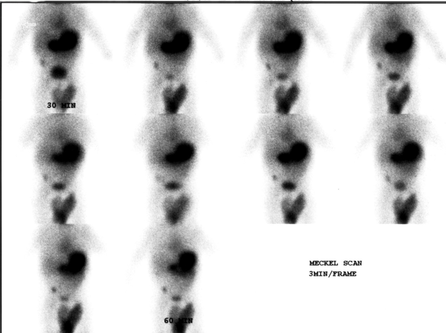 Meckel’s Diverticulum Scan with Tc-99m Pertechnetate
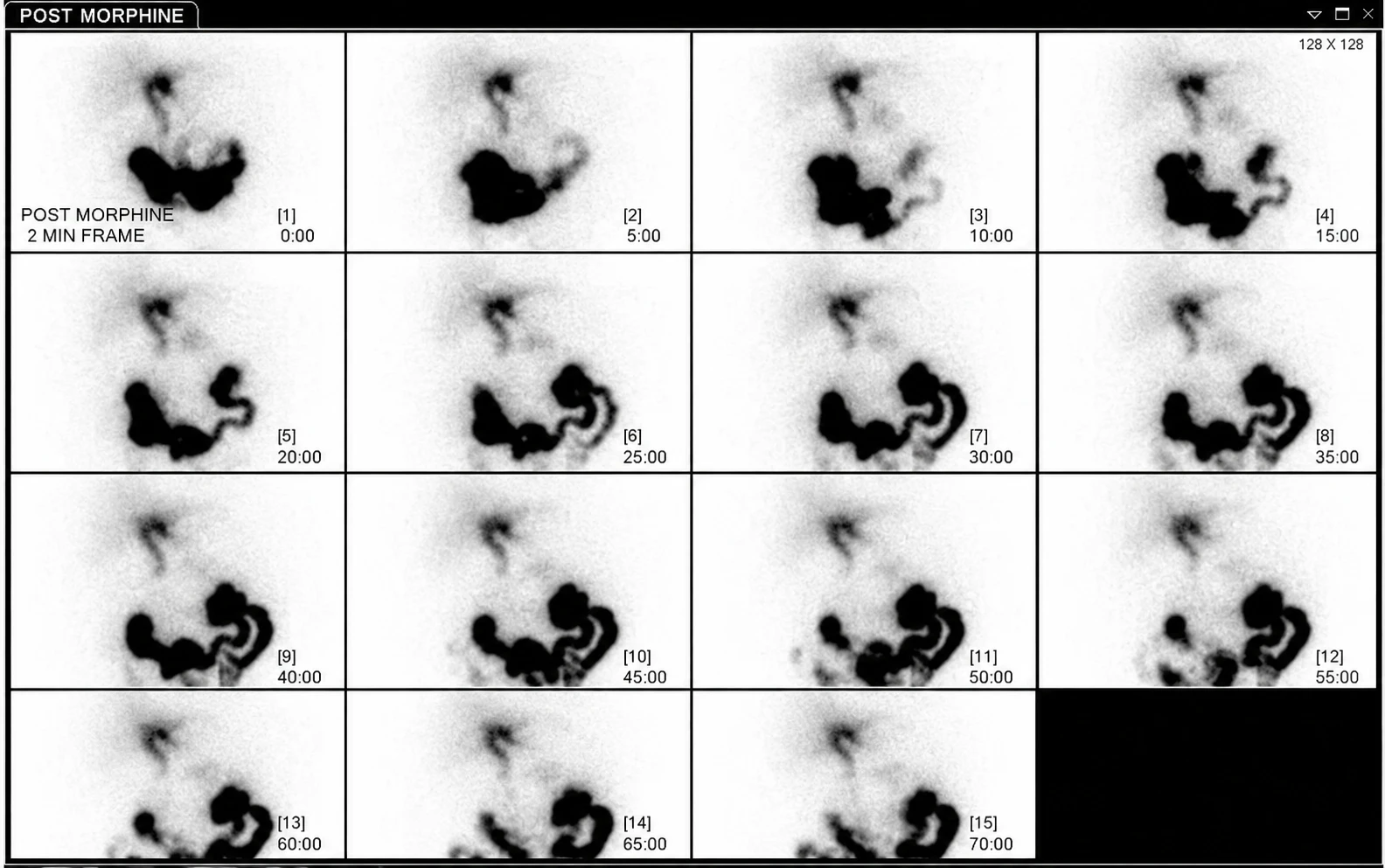 Acute Cholecystitis on HIDA Scan with Morphine
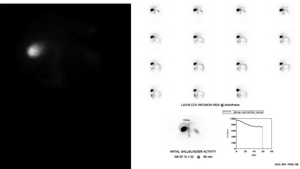 Low Gallbladder Ejection Fraction on CCK HIDA
 Biliary Atresia on Neonatal HIDA Scan
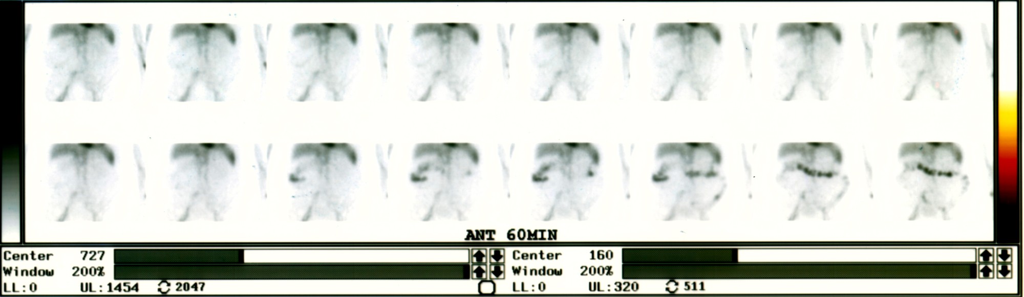 Active Lower GI Bleed on Tagged RBC Scan
All 130 nuclear medicine images by category →
Practise image interpretation in the app
All 130 images as an interactive quiz, alongside 500 NMTCB and ARRT(N) questions with worked explanations, case studies and timed mock exams. 25 questions are free, no card and no account.