Acute Cholecystitis on HIDA Scan with Morphine
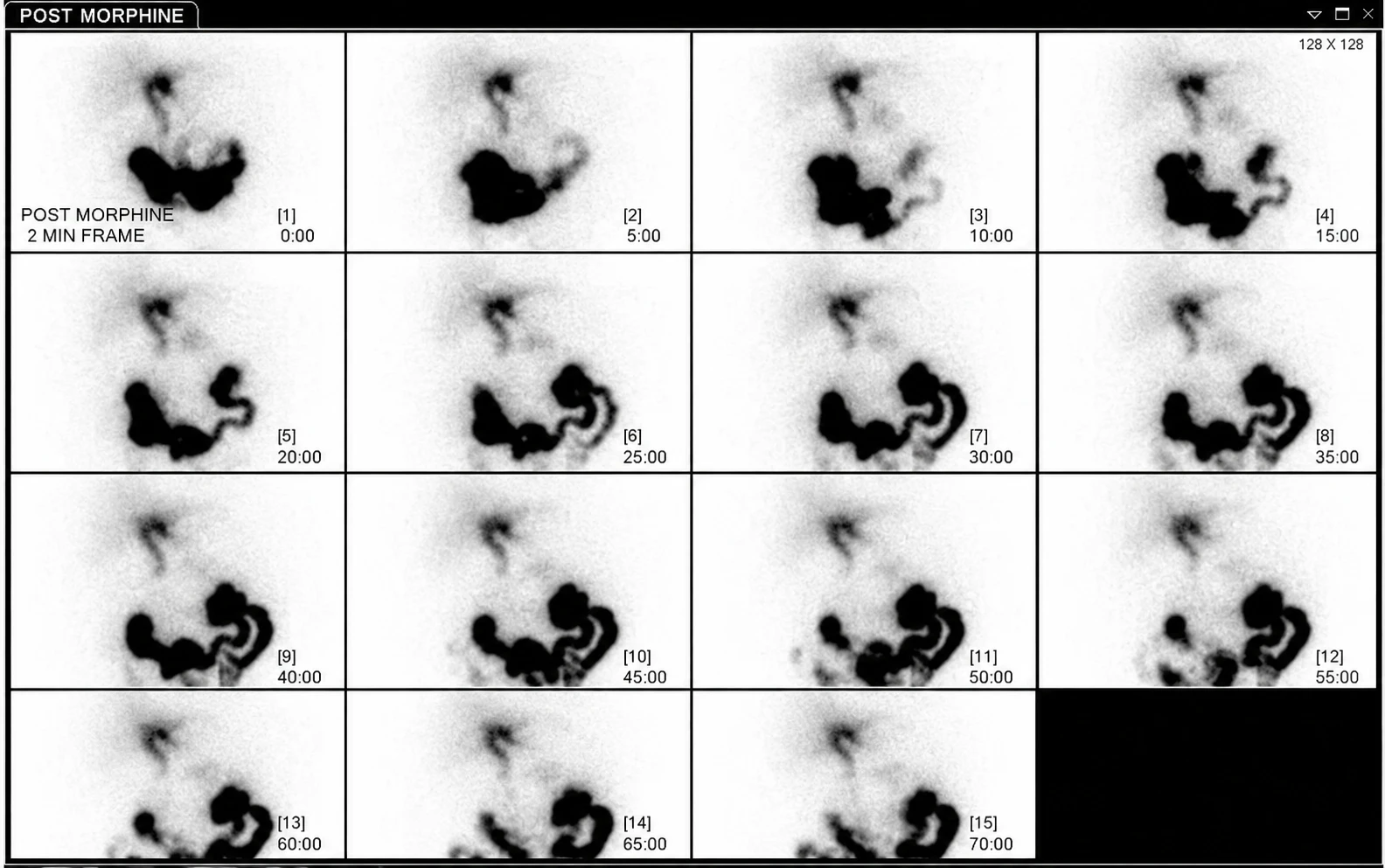
Provided clinical image
Question
A Tc-99m HIDA (hepatobiliary iminodiacetic acid) scan is performed in a patient with right upper quadrant pain and fever. The scan shows prompt hepatic uptake and biliary excretion with normal common bile duct and small bowel visualization, but NO gallbladder activity even on 4-hour delayed images. Morphine augmentation confirms persistent absent gallbladder filling. This pattern is MOST consistent with:
- AAcute cholecystitis — cystic duct obstruction prevents HIDA from entering the gallbladder
- BNormal HIDA scan
- CChronic cholecystitis
- DCommon bile duct obstruction
Show the answer and explanation
A. Acute cholecystitis — cystic duct obstruction prevents HIDA from entering the gallbladder
On a Tc-99m HIDA scan, acute cholecystitis is diagnosed by ABSENT gallbladder visualization despite good hepatic extraction and biliary excretion, due to obstruction of the cystic duct (typically by an impacted gallstone). Normally, the gallbladder fills within 30–60 minutes. Absent gallbladder activity on images up to 4 hours (with morphine augmentation), combined with normal CBD and bowel visualization, confirms cystic duct obstruction = acute cholecystitis. Sensitivity is ~96%, specificity ~90%. Morphine augmentation (increases sphincter of Oddi tone, promoting gallbladder filling if the cystic duct is patent) is used to confirm absence. False positives include prolonged fasting (>24 hrs), TPN, hepatitis.
More hepatobiliary and gi images
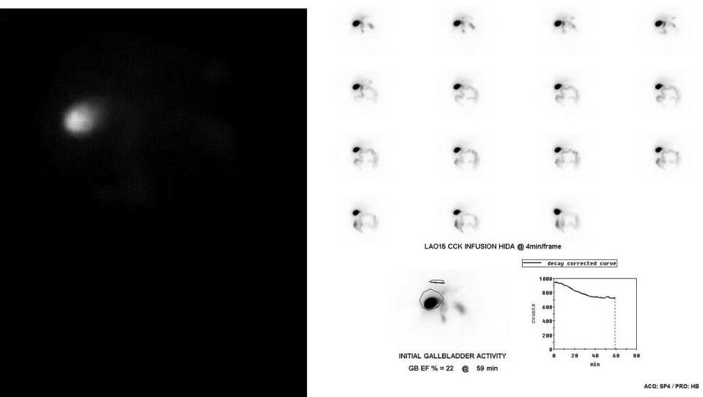 Low Gallbladder Ejection Fraction on CCK HIDA
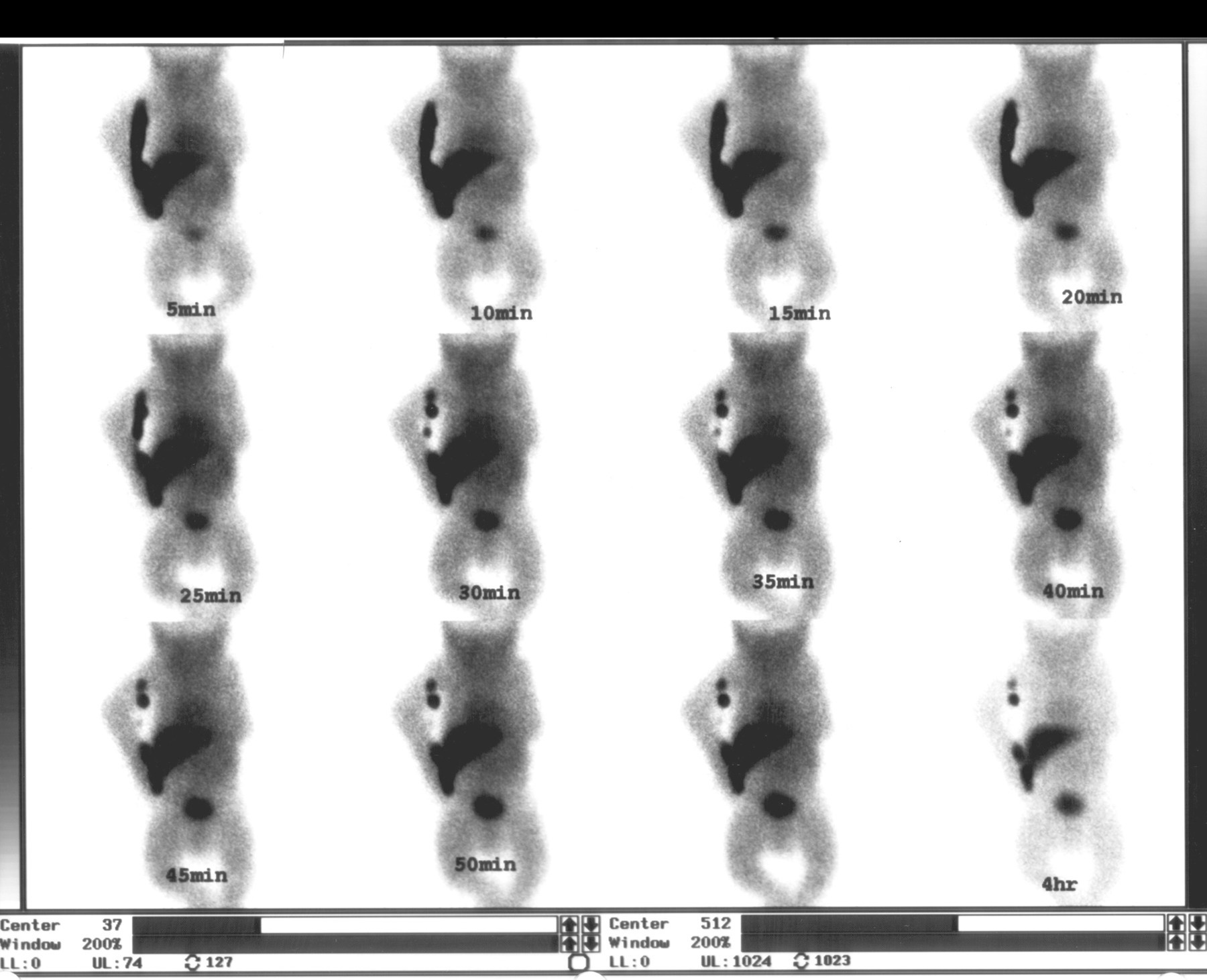 Biliary Atresia on Neonatal HIDA Scan
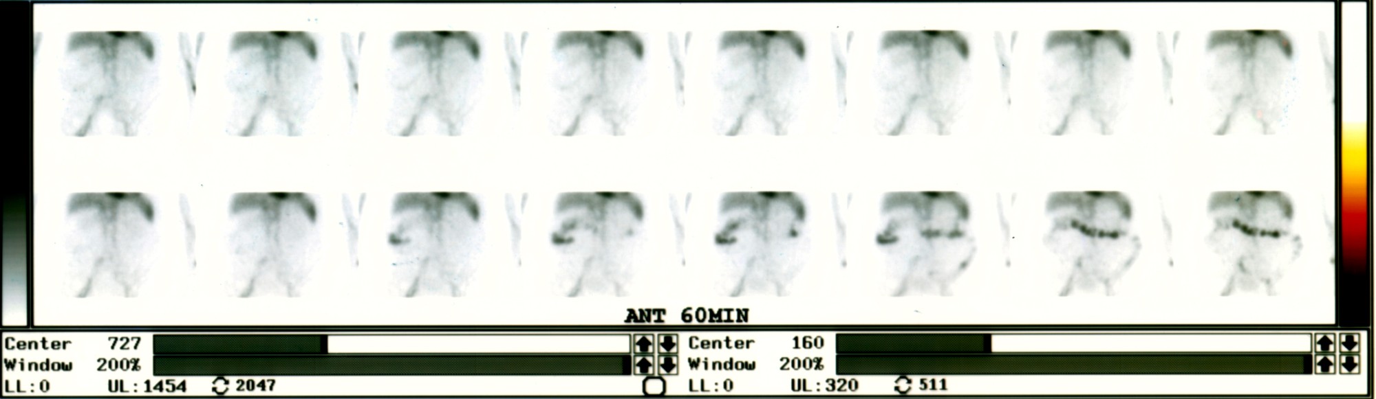 Active Lower GI Bleed on Tagged RBC Scan
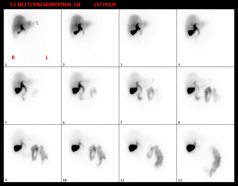 Normal Hepatobiliary Scan Sequence (Mebrofenin)
Patient Preparation for a HIDA Scan
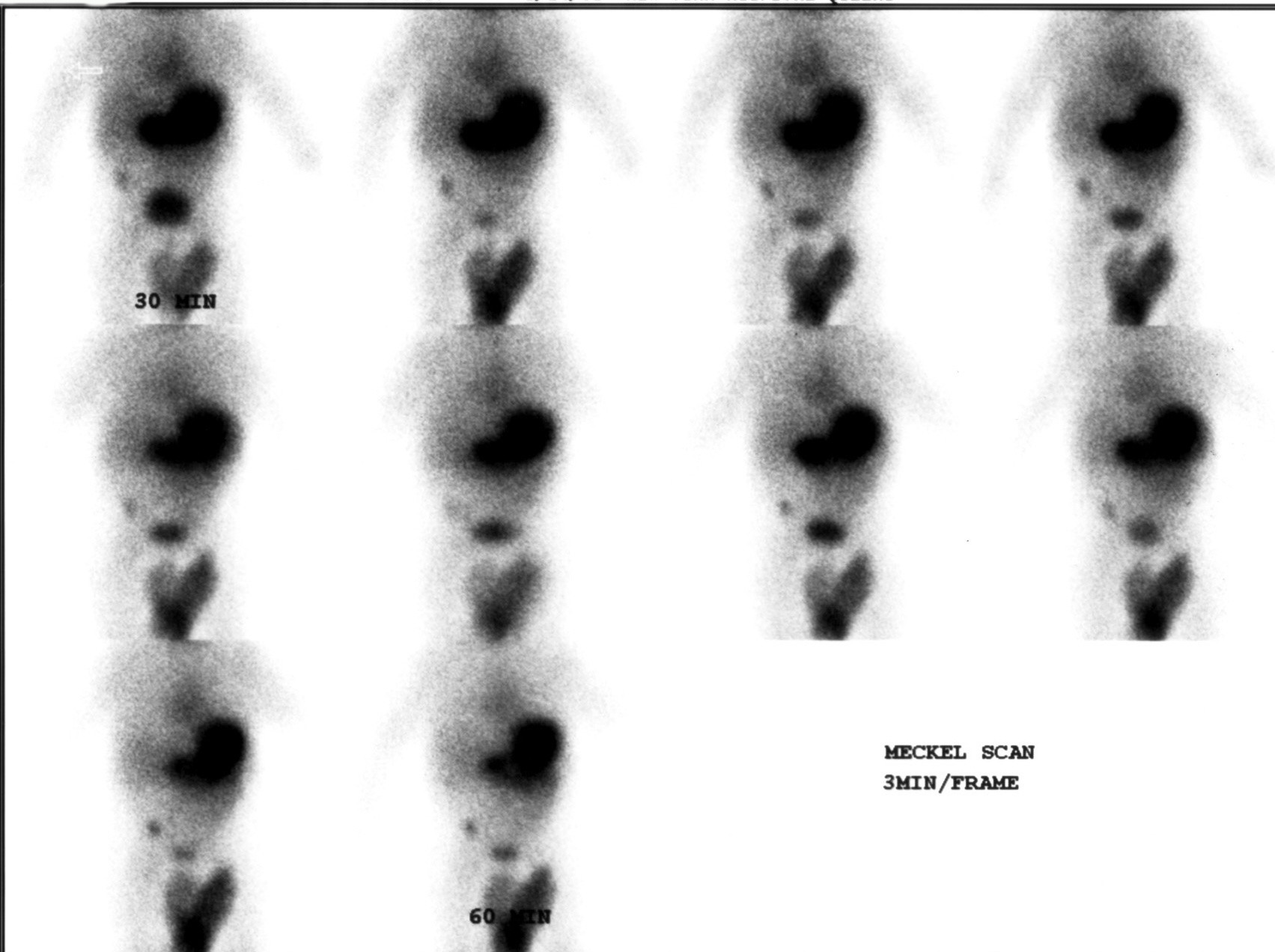 Meckel’s Diverticulum Scan with Tc-99m Pertechnetate
All 130 nuclear medicine images by category →
Practise image interpretation in the app
All 130 images as an interactive quiz, alongside 500 NMTCB and ARRT(N) questions with worked explanations, case studies and timed mock exams. 25 questions are free, no card and no account.