Meckel’s Diverticulum Scan with Tc-99m Pertechnetate
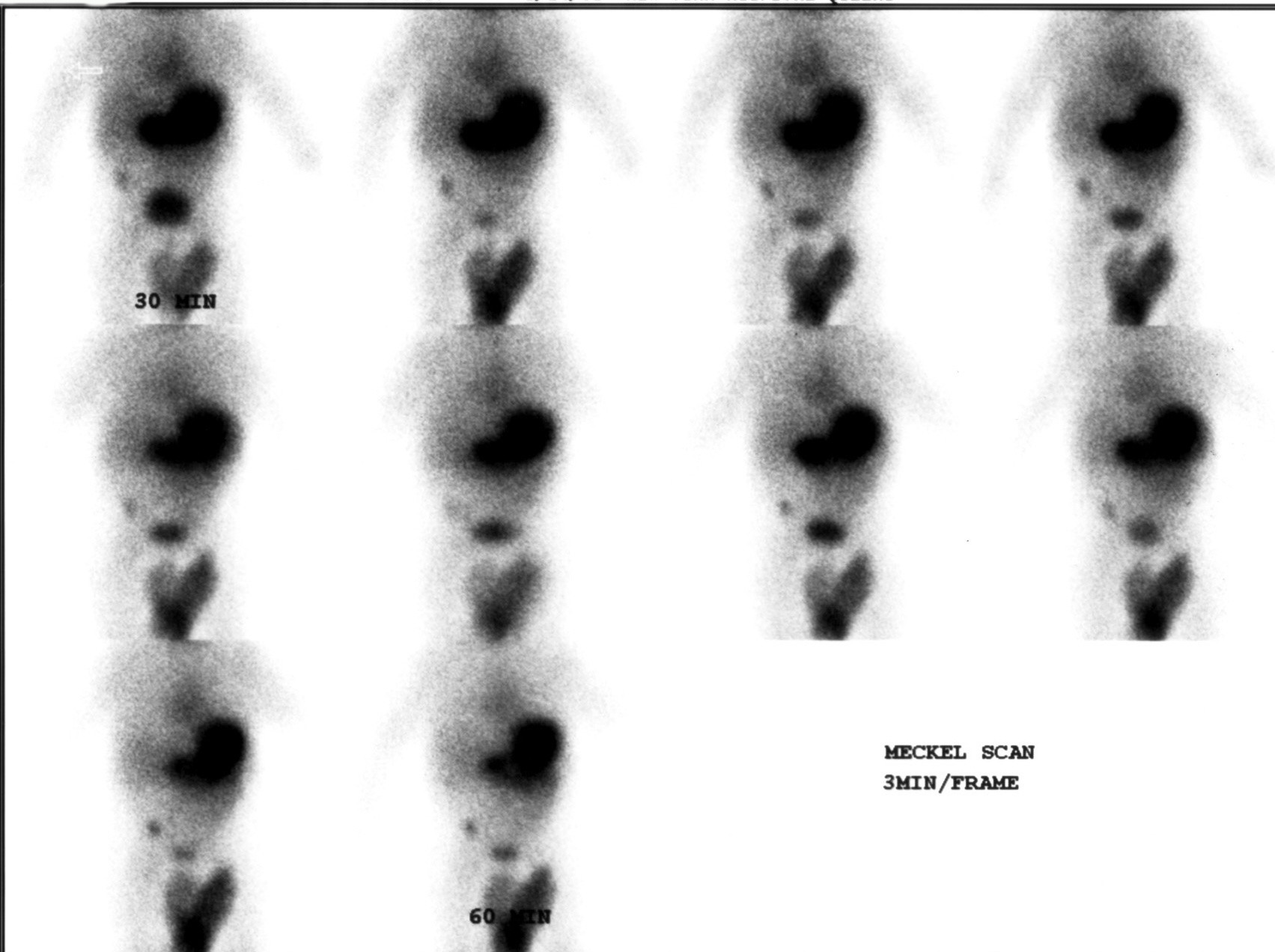
Provided clinical image
Question
In this dynamic abdominal study acquired at 3 minutes per frame, a focal area of increased activity appears in the lower abdomen at the same time as a large upper abdominal organ. Which radiopharmaceutical is used, and why does it accumulate in this abnormal location?
- ATc-99m DTPA; the tracer is filtered by the kidneys and accumulates in areas of urinary tract obstruction, causing the focal lower abdominal uptake seen in this dynamic series
- BTc-99m sulfur colloid; the tracer is taken up by reticuloendothelial cells and accumulates in ectopic liver tissue that can develop in the lower abdomen during fetal development
- CTc-99m pertechnetate; the tracer is taken up by gastric mucosa, and ectopic gastric mucosa within a Meckel's diverticulum causes the focal lower abdominal uptake seen in this study
- DTc-99m MDP; the tracer binds to areas of osteoblastic activity and accumulates in calcified soft tissue deposits that can form within abdominal diverticula
Show the answer and explanation
C. Tc-99m pertechnetate; the tracer is taken up by gastric mucosa, and ectopic gastric mucosa within a Meckel's diverticulum causes the focal lower abdominal uptake seen in this study
Tc-99m pertechnetate is used for this study. It is taken up and secreted by mucous cells of gastric mucosa. In a Meckel's diverticulum, approximately 50% of cases contain ectopic gastric mucosa, which accumulates pertechnetate just like normal stomach tissue. On a positive scan, focal uptake appears in the lower abdomen (typically right lower quadrant) simultaneously with stomach uptake and persists throughout the dynamic series. The study is acquired at 3 min/frame for 45–60 minutes.
More hepatobiliary and gi images
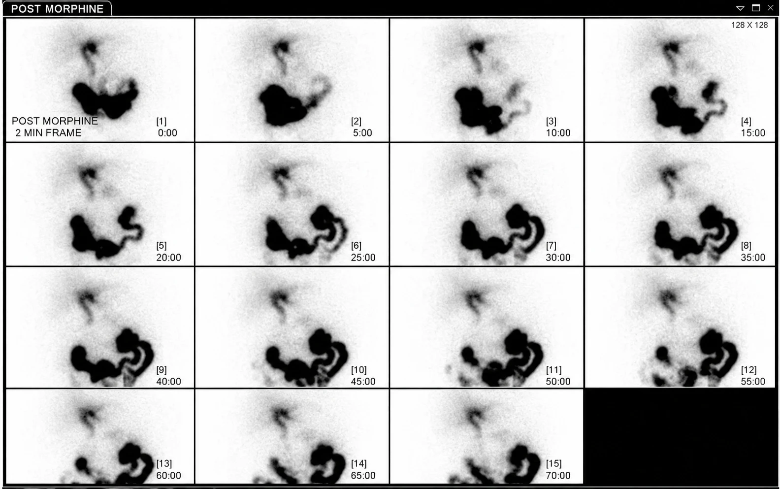 Acute Cholecystitis on HIDA Scan with Morphine
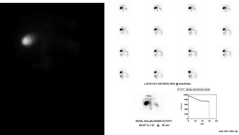 Low Gallbladder Ejection Fraction on CCK HIDA
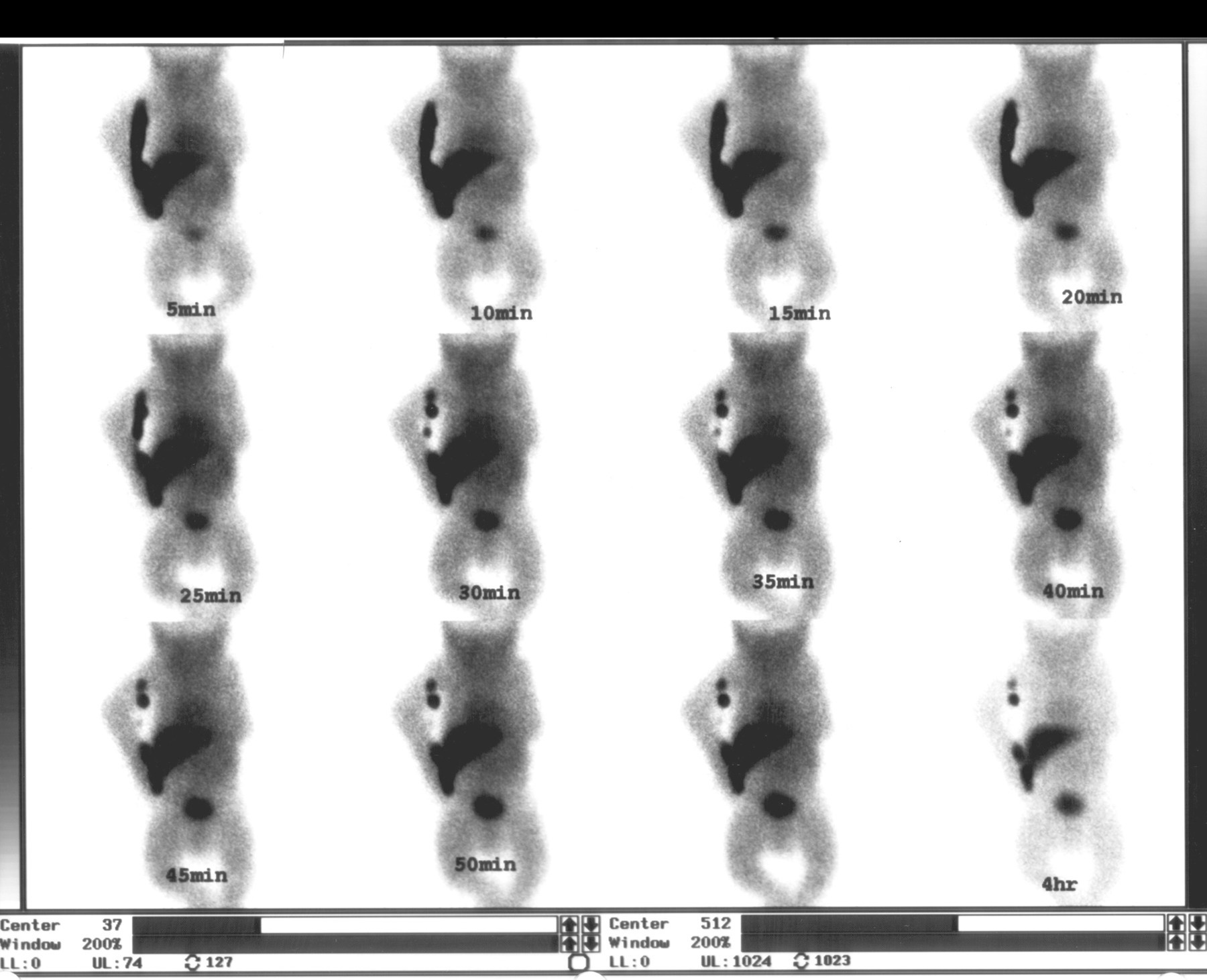 Biliary Atresia on Neonatal HIDA Scan
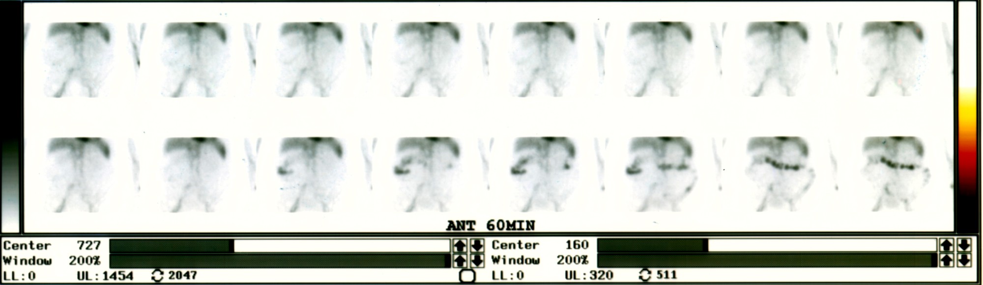 Active Lower GI Bleed on Tagged RBC Scan
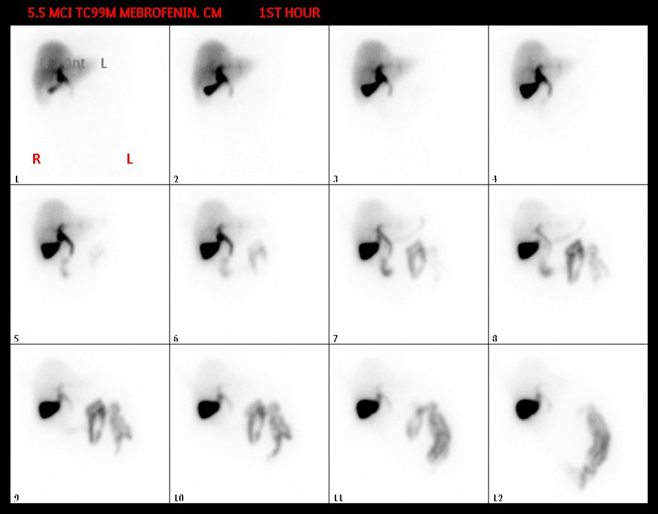 Normal Hepatobiliary Scan Sequence (Mebrofenin)
Patient Preparation for a HIDA Scan
All 130 nuclear medicine images by category →
Practise image interpretation in the app
All 130 images as an interactive quiz, alongside 500 NMTCB and ARRT(N) questions with worked explanations, case studies and timed mock exams. 25 questions are free, no card and no account.