Active Lower GI Bleed on Tagged RBC Scan
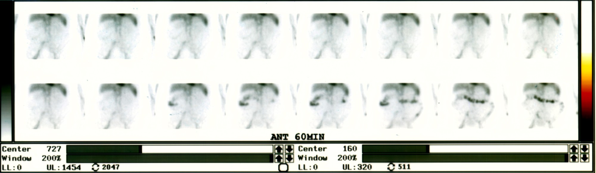
Provided clinical image
Question
A Tc-99m red blood cell (RBC) scan is performed in a patient with hematochezia and a hemoglobin drop. The scan shows a focal area of activity in the right lower quadrant that INCREASES and MOVES (migrates) on sequential images. This pattern is MOST consistent with:
- AHepatic hemangioma — stable RBC uptake in the liver
- BArtifact from patient motion
- CActive lower GI bleeding in the right colon — activity appears, increases, and moves along the bowel lumen as bleeding progresses
- DMeckel's diverticulum
Show the answer and explanation
C. Active lower GI bleeding in the right colon — activity appears, increases, and moves along the bowel lumen as bleeding progresses
Tc-99m RBC scanning is used to localize active GI bleeding. The key feature of a true bleed is: (1) NEW focal activity appearing where none was before (vs. physiologic activity in heart, great vessels, liver, spleen, kidneys, bladder); (2) INCREASING activity over time; (3) MIGRATION along the bowel lumen in the direction of peristalsis. This dynamic pattern distinguishes true hemorrhage from hepatic hemangioma (stable, non-migratory) or background activity. Tc-99m RBC scan can detect bleeding rates as low as 0.04–0.1 mL/min, making it more sensitive than angiography (0.5–1 mL/min) for intermittent bleeds. Positive scans guide selective angiography and hemostasis.
More hepatobiliary and gi images
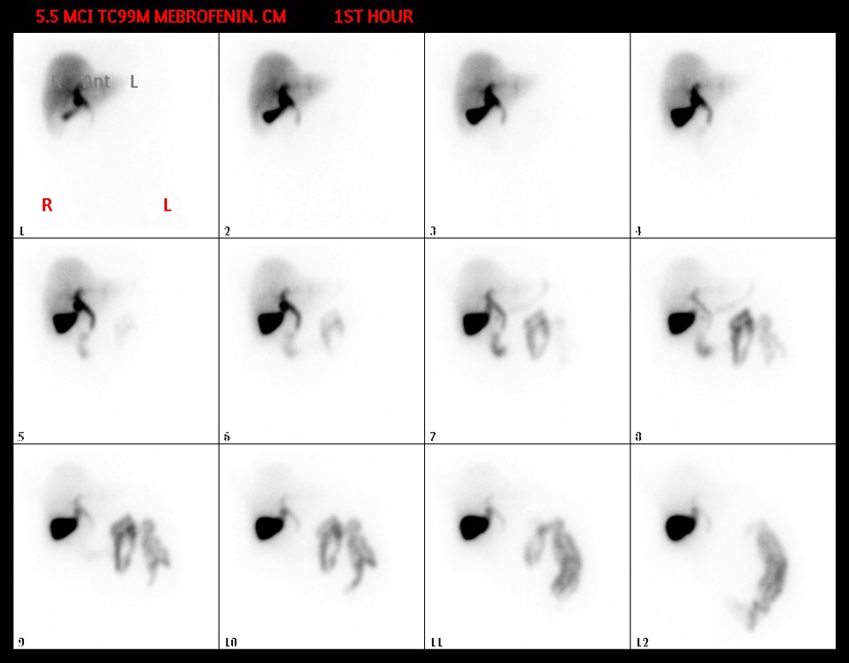 Normal Hepatobiliary Scan Sequence (Mebrofenin)
Patient Preparation for a HIDA Scan
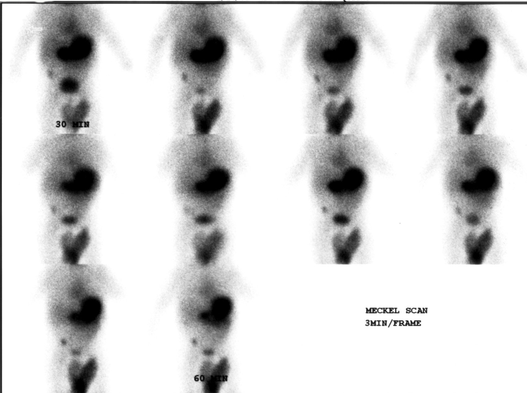 Meckel’s Diverticulum Scan with Tc-99m Pertechnetate
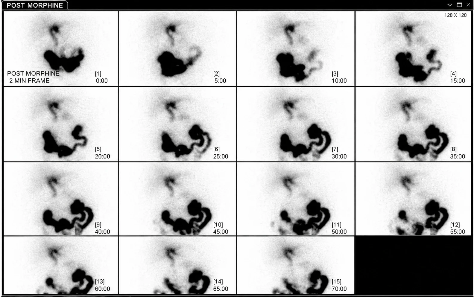 Acute Cholecystitis on HIDA Scan with Morphine
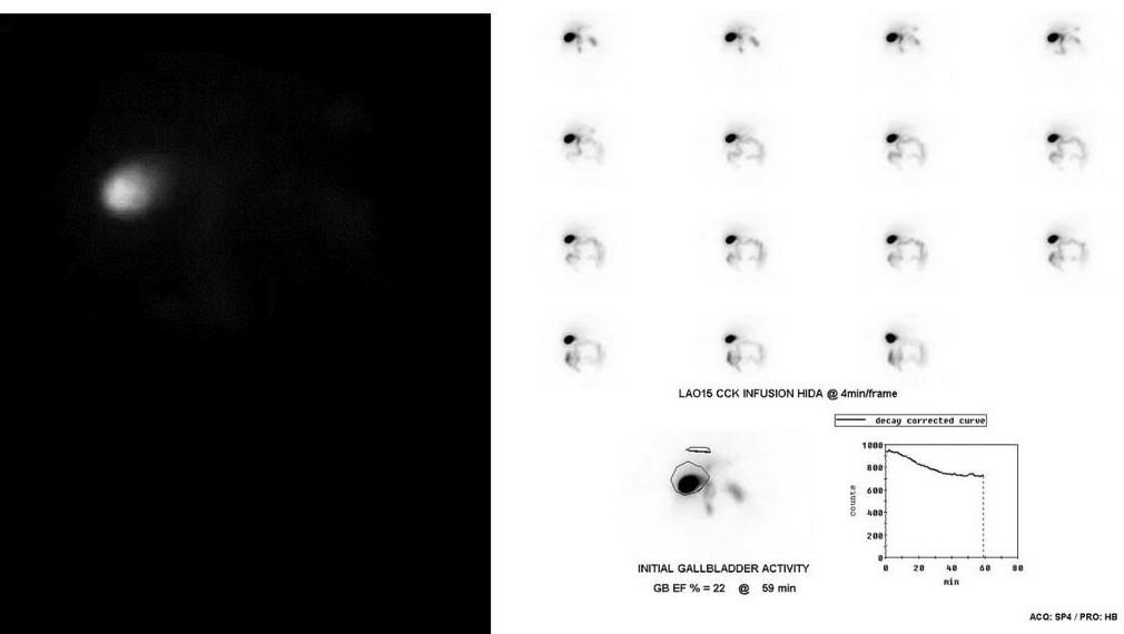 Low Gallbladder Ejection Fraction on CCK HIDA
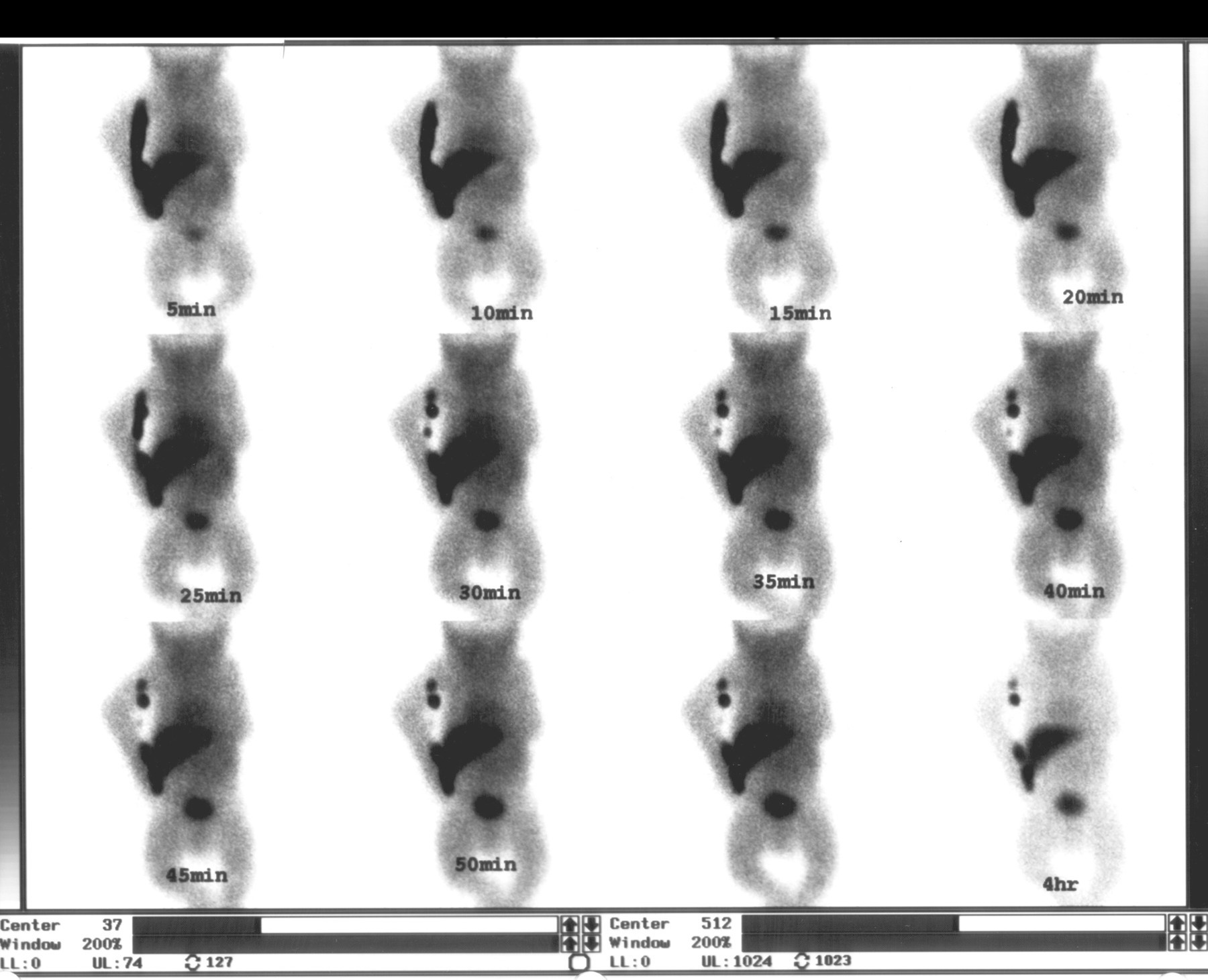 Biliary Atresia on Neonatal HIDA Scan
All 130 nuclear medicine images by category →
Practise image interpretation in the app
All 130 images as an interactive quiz, alongside 500 NMTCB and ARRT(N) questions with worked explanations, case studies and timed mock exams. 25 questions are free, no card and no account.