Low Gallbladder Ejection Fraction on CCK HIDA
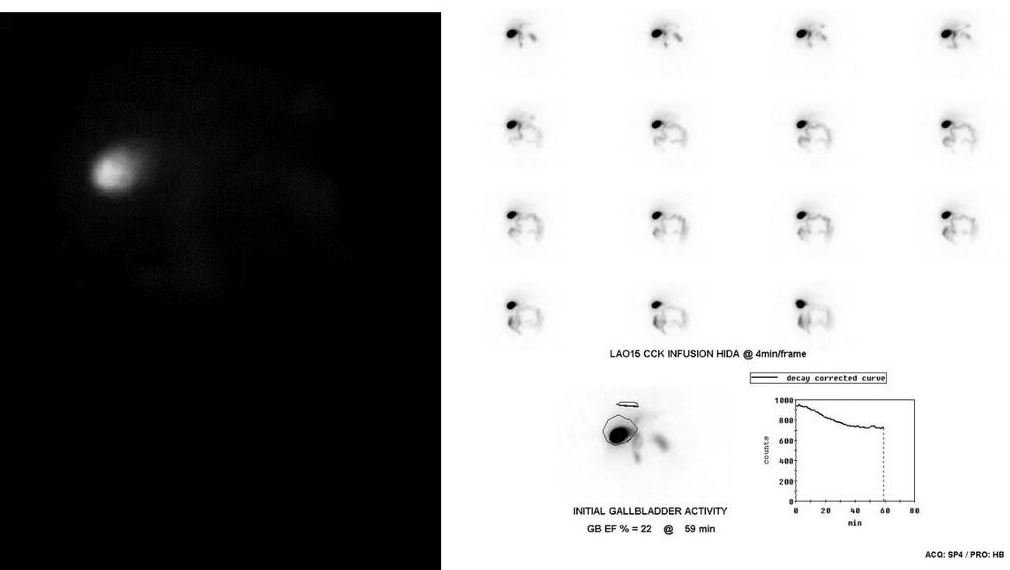
Provided clinical image
Question
A Tc-99m HIDA scan with CCK (cholecystokinin) stimulation is performed in a patient with chronic right upper quadrant pain. The gallbladder fills normally but after CCK infusion, the gallbladder ejection fraction (GBEF) is calculated at 22% (normal >35%). This finding is MOST consistent with:
- AAcute cholecystitis — cystic duct obstruction by an impacted calculus causing gallbladder wall edema, pericholecystic fluid, and absent filling on standard HIDA imaging without requiring CCK provocation
- BChronic cholecystitis (biliary dyskinesia) — impaired gallbladder contractility with reduced GBEF
- CCommon bile duct obstruction — elevated biliary backpressure causing uniform dilatation of the biliary tree with prolonged tracer transit and paradoxical gallbladder hyperconcentration due to increased absorption time
- DNormal gallbladder function — GBEF values between 20–35% represent a borderline physiologic range attributable to CCK infusion rate variability, fasting duration, or interobserver ROI placement differences
Show the answer and explanation
B. Chronic cholecystitis (biliary dyskinesia) — impaired gallbladder contractility with reduced GBEF
Gallbladder ejection fraction (GBEF) is measured on CCK-HIDA scan by calculating the percentage of gallbladder activity ejected after CCK infusion: GBEF = [(Maximum GB counts – Minimum GB counts) / Maximum GB counts] × 100%. Normal GBEF is >35% (some use >38% or >40%). A reduced GBEF (<35%) indicates impaired gallbladder contractility, consistent with chronic cholecystitis / biliary dyskinesia. This diagnosis explains chronic RUQ pain in patients with normal ultrasound (no stones). Cholecystectomy in patients with reduced GBEF and typical biliary-type pain relieves symptoms in ~90% of cases. The GBEF can also be low in prolonged fasting, TPN, or opioid use.
More hepatobiliary and gi images
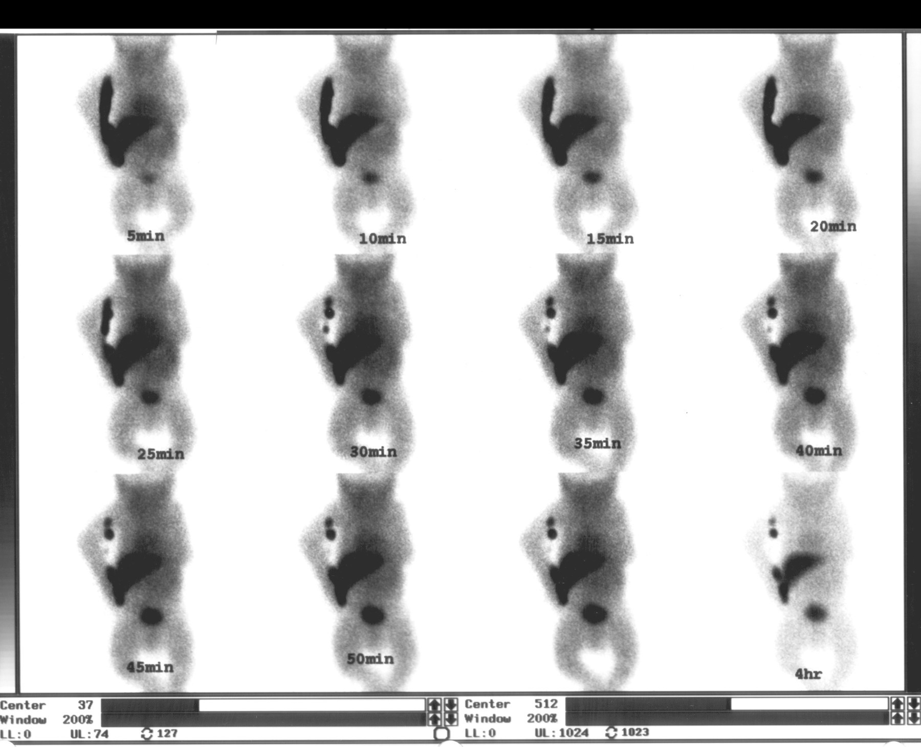 Biliary Atresia on Neonatal HIDA Scan
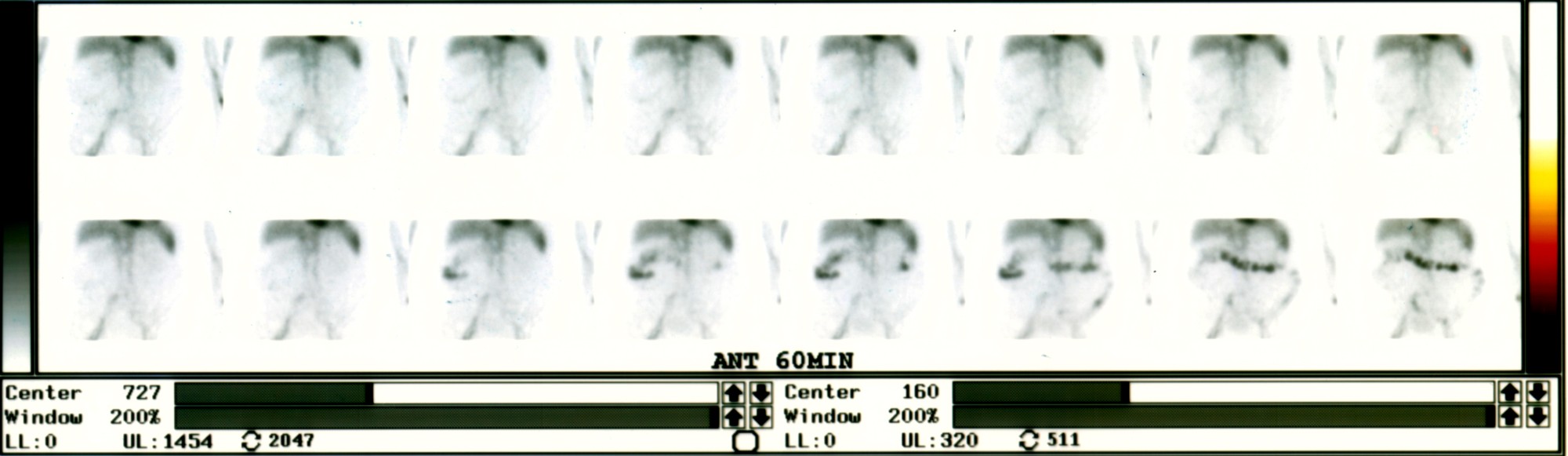 Active Lower GI Bleed on Tagged RBC Scan
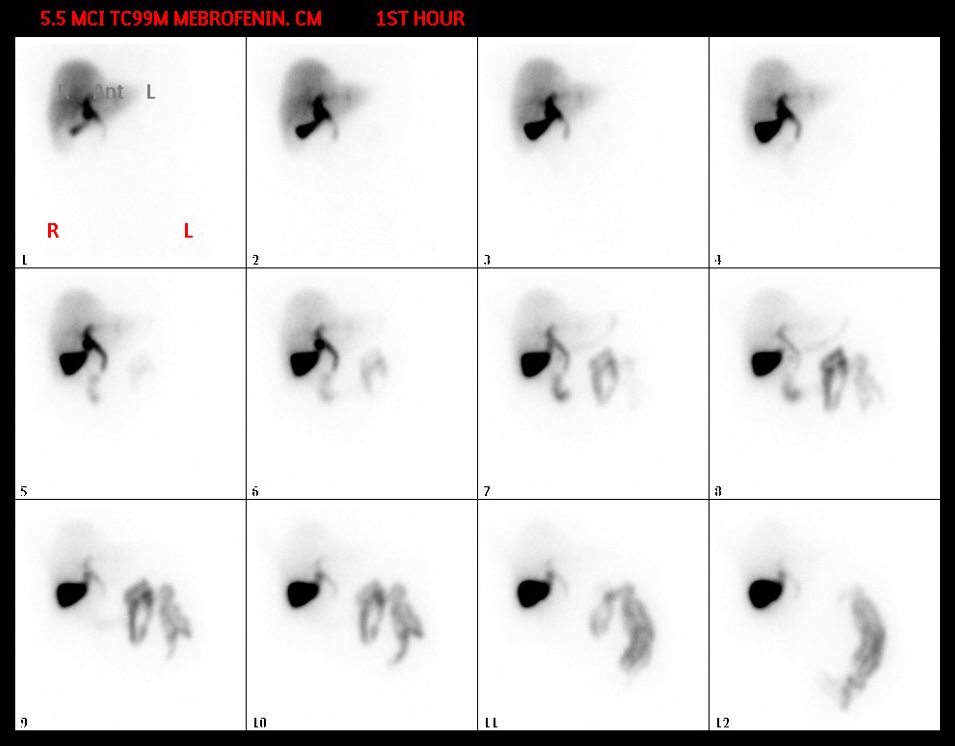 Normal Hepatobiliary Scan Sequence (Mebrofenin)
Patient Preparation for a HIDA Scan
 Meckel’s Diverticulum Scan with Tc-99m Pertechnetate
 Acute Cholecystitis on HIDA Scan with Morphine
All 130 nuclear medicine images by category →
Practise image interpretation in the app
All 130 images as an interactive quiz, alongside 500 NMTCB and ARRT(N) questions with worked explanations, case studies and timed mock exams. 25 questions are free, no card and no account.