Parathyroid Adenoma on Dual-Phase Sestamibi Scan
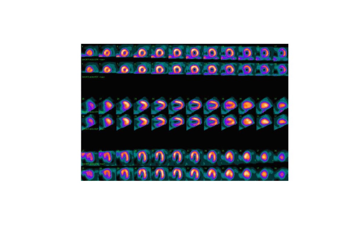
CC BY-SA 4.0 · Sincefalastrum / Wikimedia Commons · source file
Question
Tc-99m sestamibi dual-phase parathyroid scintigraphy is performed in a patient with primary hyperparathyroidism (elevated PTH and calcium). Early images show uptake in both the thyroid and a right inferior parathyroid region; delayed images (2 hours) show PERSISTENT uptake in the right inferior parathyroid region with washout from the thyroid. This pattern is MOST consistent with:
- ANormal parathyroid glands
- BThyroid adenoma
- CParathyroid carcinoma — always shows washout
- DRight inferior parathyroid adenoma — persistent sestamibi retention on delayed images indicates hyperfunctioning parathyroid tissue
Show the answer and explanation
D. Right inferior parathyroid adenoma — persistent sestamibi retention on delayed images indicates hyperfunctioning parathyroid tissue
Tc-99m sestamibi dual-phase parathyroid imaging exploits differential washout kinetics: normal thyroid tissue and normal parathyroid glands wash out sestamibi quickly, while hyperfunctioning parathyroid adenomas (which have abundant mitochondria from hyperplastic chief cells) RETAIN sestamibi on delayed images. The characteristic pattern of persistent focal uptake on 2-hour delayed images (with thyroid washout) localizes the parathyroid adenoma. Subtraction technique (Tc-99m sestamibi minus Tc-99m pertechnetate) further improves localization. SPECT/CT improves 3D localization for surgical planning. Sensitivity for single adenoma is ~85–90%; lower for hyperplasia or double adenomas.
More thyroid and parathyroid images
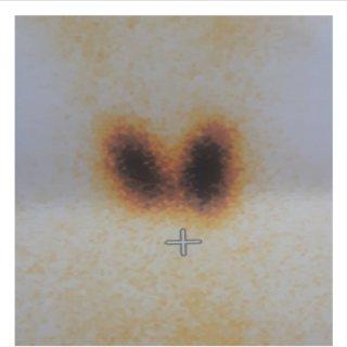 Graves’ Disease on Thyroid Scan
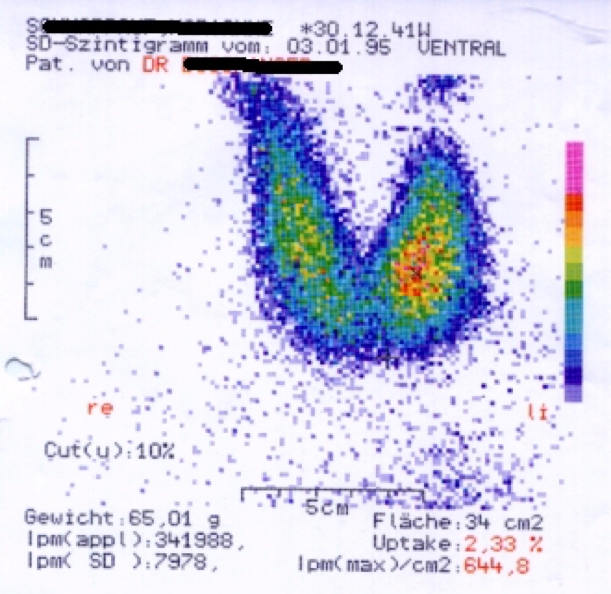 Hot Thyroid Nodule: Autonomous Function
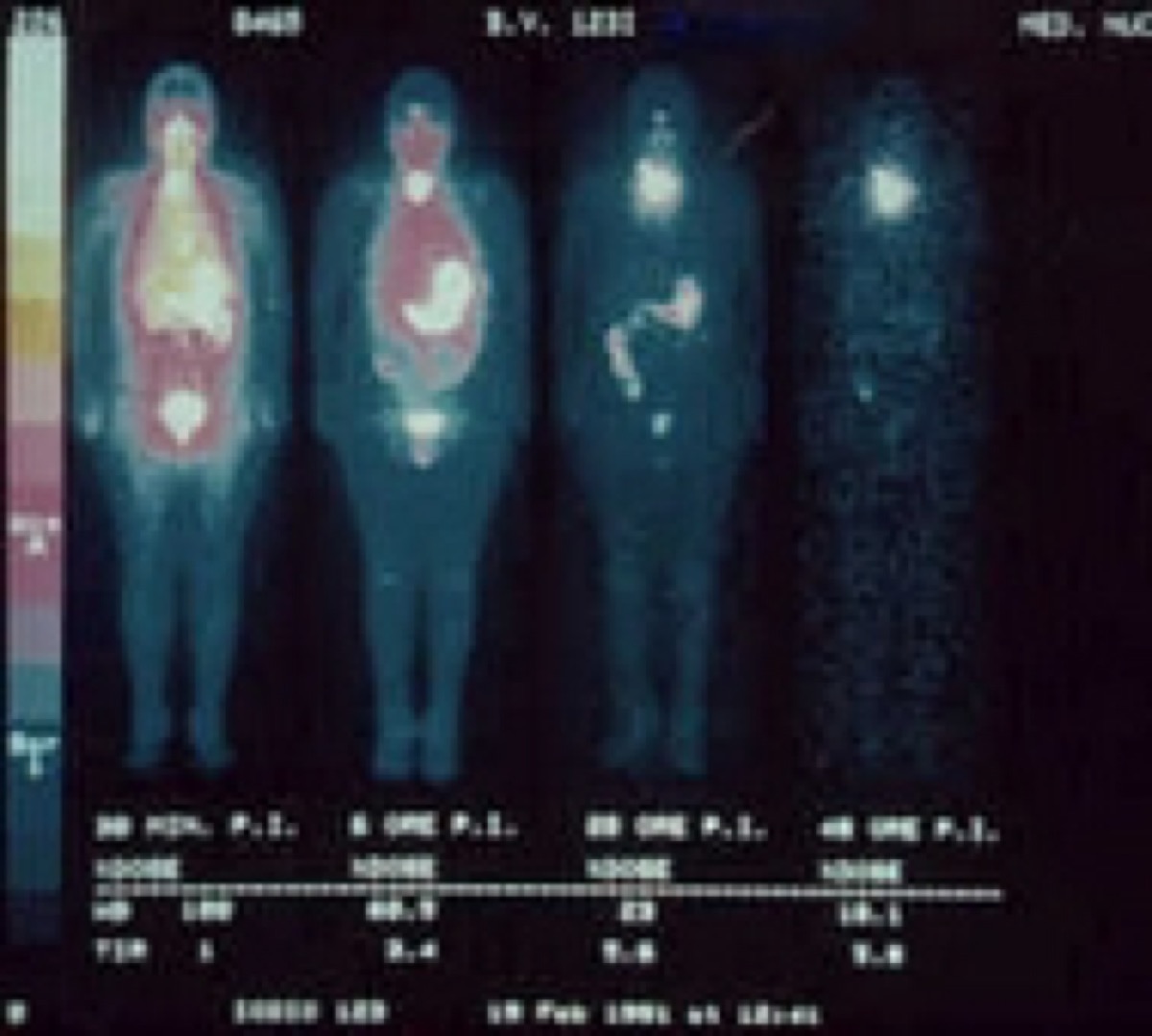 Why I-123 Is Used for Diagnostic Thyroid Imaging
I-131 vs I-123 for Post-Thyroidectomy Whole-Body Scans
Cold Thyroid Nodule: The Next Step
Lingual Thyroid on I-123 Scan
All 130 nuclear medicine images by category →
Practise image interpretation in the app
All 130 images as an interactive quiz, alongside 500 NMTCB and ARRT(N) questions with worked explanations, case studies and timed mock exams. 25 questions are free, no card and no account.