In-111 Oxine WBC Scan: Agent and Timing
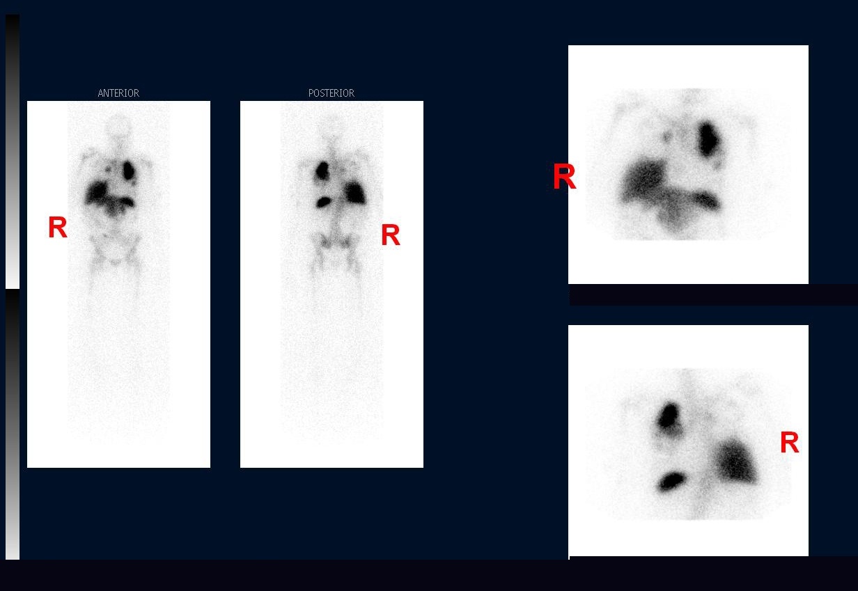
Provided clinical image
Question
This whole-body scan shows anterior and posterior views along with abdominal spot images. Based on the biodistribution pattern — prominent uptake in the liver, spleen, and abnormal lung activity — which radiopharmaceutical was most likely used and what is its standard imaging schedule?
- ATc-99m HMPAO-labeled WBCs; images are obtained at 1, 4, and 24 hours after reinjection, with delayed imaging used to improve specificity for chronic infection
- BIn-111 oxine-labeled WBCs; images are typically obtained at 24 hours after reinjection, with an optional earlier image at 4 hours post-reinjection
- CGa-67 citrate; the radiopharmaceutical is injected directly without any in vitro cell labeling, and images are acquired at 24, 48, and 72 hours
- DTc-99m sulfur colloid; images are obtained 30–60 minutes after reinjection to assess white blood cell distribution in the liver, spleen, and bone marrow
Show the answer and explanation
B. In-111 oxine-labeled WBCs; images are typically obtained at 24 hours after reinjection, with an optional earlier image at 4 hours post-reinjection
In-111 oxine (indium-111 oxyquinoline) is used to label white blood cells in vitro: the patient's blood is drawn, WBCs are isolated, incubated with In-111 oxine, then reinjected. Images are typically acquired at 24 hours post-reinjection to allow adequate WBC migration to sites of infection, with an optional 4-hour early image. The physical half-life of In-111 (67.3 hours) makes 24-hour delayed imaging practical.
More infection and inflammation images
Abnormal Lung Uptake on In-111 WBC Scan
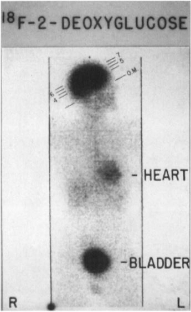 FDG PET/CT for Fever of Unknown Origin
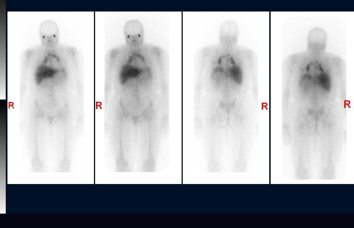 Sarcoidosis on Gallium Scan: Panda and Lambda Signs
Gallium-67 Imaging Times for Sarcoidosis
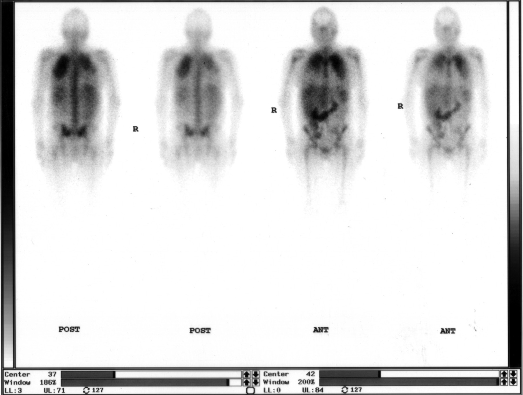 Ga-67 Citrate Whole-Body Scan: Agent and Timing
Abnormal Bilateral Lung Uptake on Gallium Scan
All 130 nuclear medicine images by category →
Practise image interpretation in the app
All 130 images as an interactive quiz, alongside 500 NMTCB and ARRT(N) questions with worked explanations, case studies and timed mock exams. 25 questions are free, no card and no account.