Abnormal Lung Uptake on In-111 WBC Scan
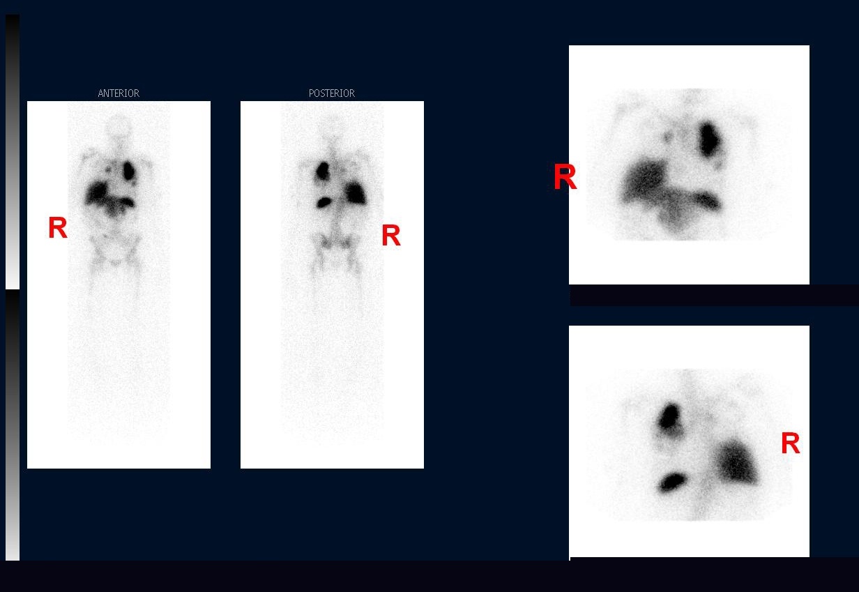
Provided clinical image
Question
On this In-111 WBC scan, the lungs show prominent bilateral uptake. Why is this finding considered abnormal on delayed images?
- AThe lungs always show high WBC uptake on In-111 studies because labeled cells are cleared by pulmonary capillaries, making any lung activity an expected and clinically insignificant finding
- BLung activity indicates the labeling procedure was unsuccessful, as properly labeled cells accumulate only in the blood pool and should not localize in any organ at 24 hours
- CProminent lung uptake is a known scatter artifact caused by the energy of In-111, which creates apparent pulmonary activity from adjacent high-uptake organs like the liver and spleen
- DAfter the first few hours, labeled WBCs normally distribute to the liver, spleen, and bone marrow; persistent lung uptake on delayed images is abnormal and suggests active pulmonary infection or inflammation
Show the answer and explanation
D. After the first few hours, labeled WBCs normally distribute to the liver, spleen, and bone marrow; persistent lung uptake on delayed images is abnormal and suggests active pulmonary infection or inflammation
Normal In-111 WBC biodistribution on delayed (24-hour) images is liver > spleen > bone marrow. Early transient lung uptake can be seen within the first hour due to WBC transit through the pulmonary vasculature, but this should clear. Persistent or prominent bilateral lung activity on delayed images is abnormal and indicates an active pulmonary process such as pneumonia, ARDS, or diffuse pulmonary infection.
More infection and inflammation images
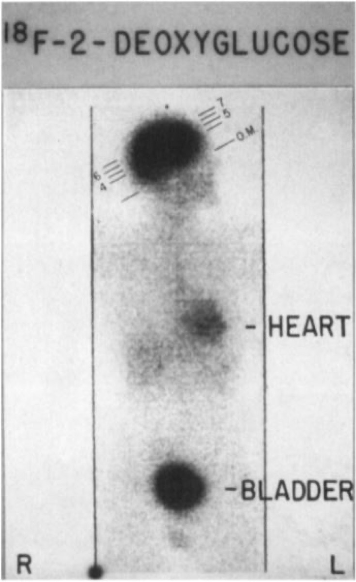 FDG PET/CT for Fever of Unknown Origin
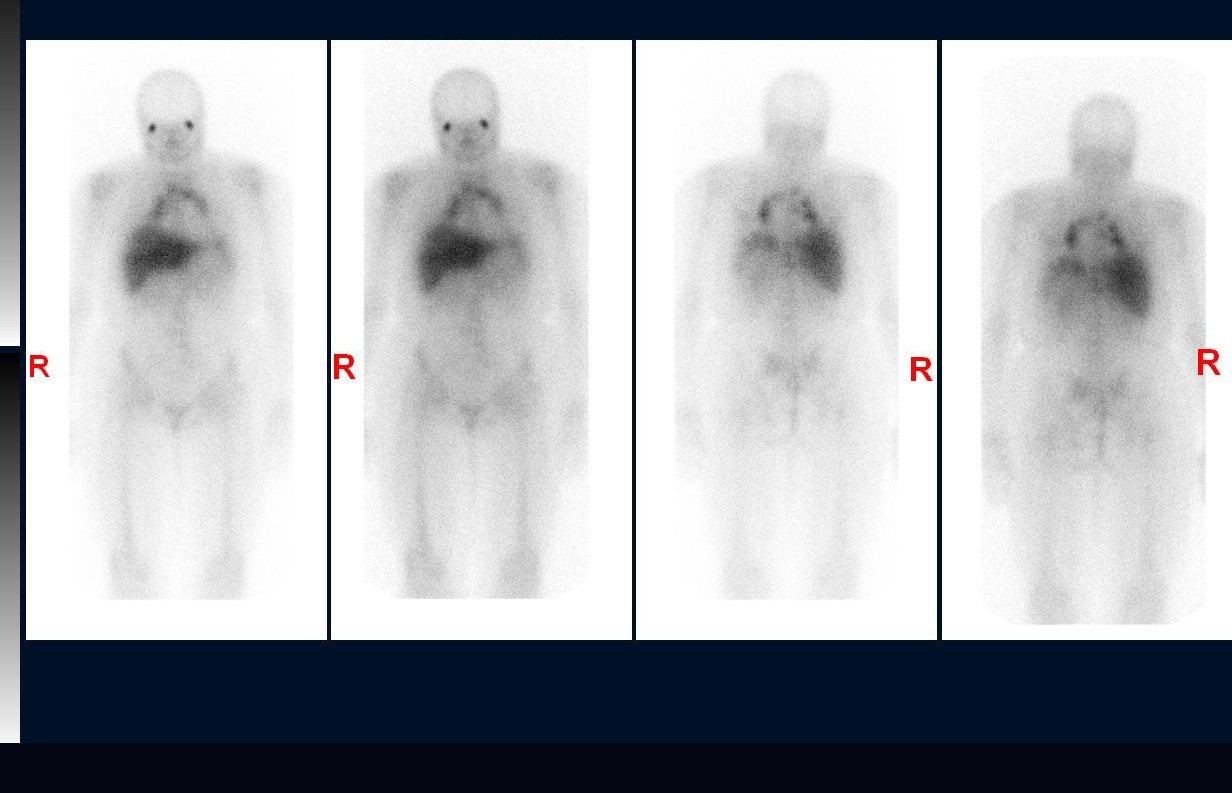 Sarcoidosis on Gallium Scan: Panda and Lambda Signs
Gallium-67 Imaging Times for Sarcoidosis
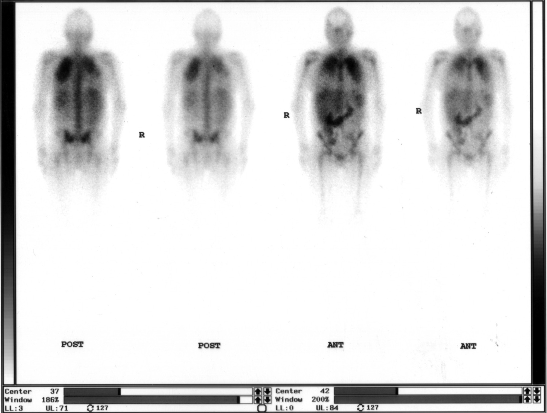 Ga-67 Citrate Whole-Body Scan: Agent and Timing
Abnormal Bilateral Lung Uptake on Gallium Scan
In-111 Oxine WBC Scan: Agent and Timing
All 130 nuclear medicine images by category →
Practise image interpretation in the app
All 130 images as an interactive quiz, alongside 500 NMTCB and ARRT(N) questions with worked explanations, case studies and timed mock exams. 25 questions are free, no card and no account.