FDG PET/CT for Fever of Unknown Origin
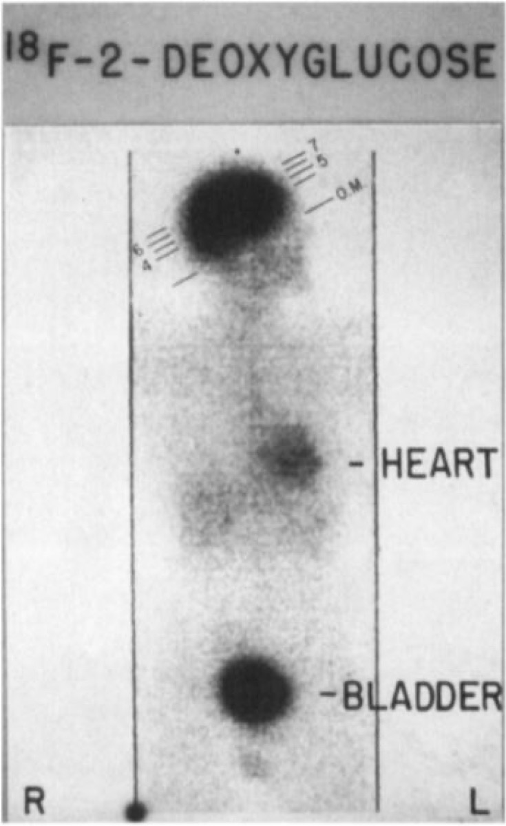
CC BY-SA 4.0 · Pennradiology / Wikimedia Commons · source file
Question
A patient has a 3-week history of fever >38.3°C, weight loss, and night sweats without an identified source despite extensive workup (blood cultures, CT, echocardiography). FDG PET/CT is ordered for evaluation of fever of unknown origin (FUO). What is the PRIMARY value of FDG PET/CT in this clinical setting?
- AFDG PET is always negative in infection, confirming non-infectious causes
- BFDG PET provides a radiation-free alternative to CT
- CFDG PET/CT localizes occult infection, inflammation, or malignancy in approximately 50–60% of FUO cases, directing further targeted evaluation or biopsy
- DFDG PET replaces blood cultures as the gold standard for FUO
Show the answer and explanation
C. FDG PET/CT localizes occult infection, inflammation, or malignancy in approximately 50–60% of FUO cases, directing further targeted evaluation or biopsy
FDG PET/CT has emerged as an important diagnostic tool for fever of unknown origin (FUO) because many causes — infection (endocarditis, osteomyelitis, abscesses), vasculitis (large-vessel vasculitis such as giant cell arteritis), inflammatory conditions, and occult malignancy — show increased FDG uptake. Meta-analyses show FDG PET/CT identifies a potential diagnostic focus in ~50–60% of FUO cases, guiding further targeted evaluation or biopsy. This makes it one of the highest-yield single studies in the FUO workup algorithm. It is particularly valuable for detecting large-vessel vasculitis and lymphoma that may be missed by CT alone.
More infection and inflammation images
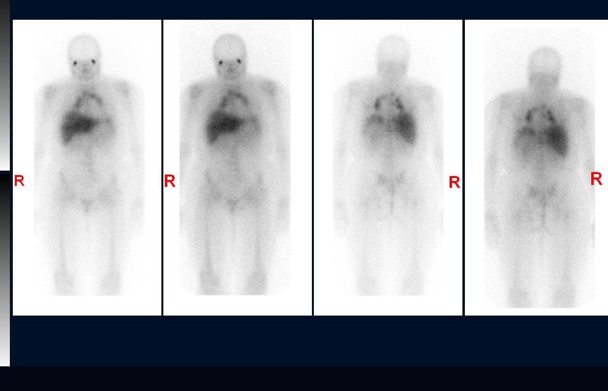 Sarcoidosis on Gallium Scan: Panda and Lambda Signs
Gallium-67 Imaging Times for Sarcoidosis
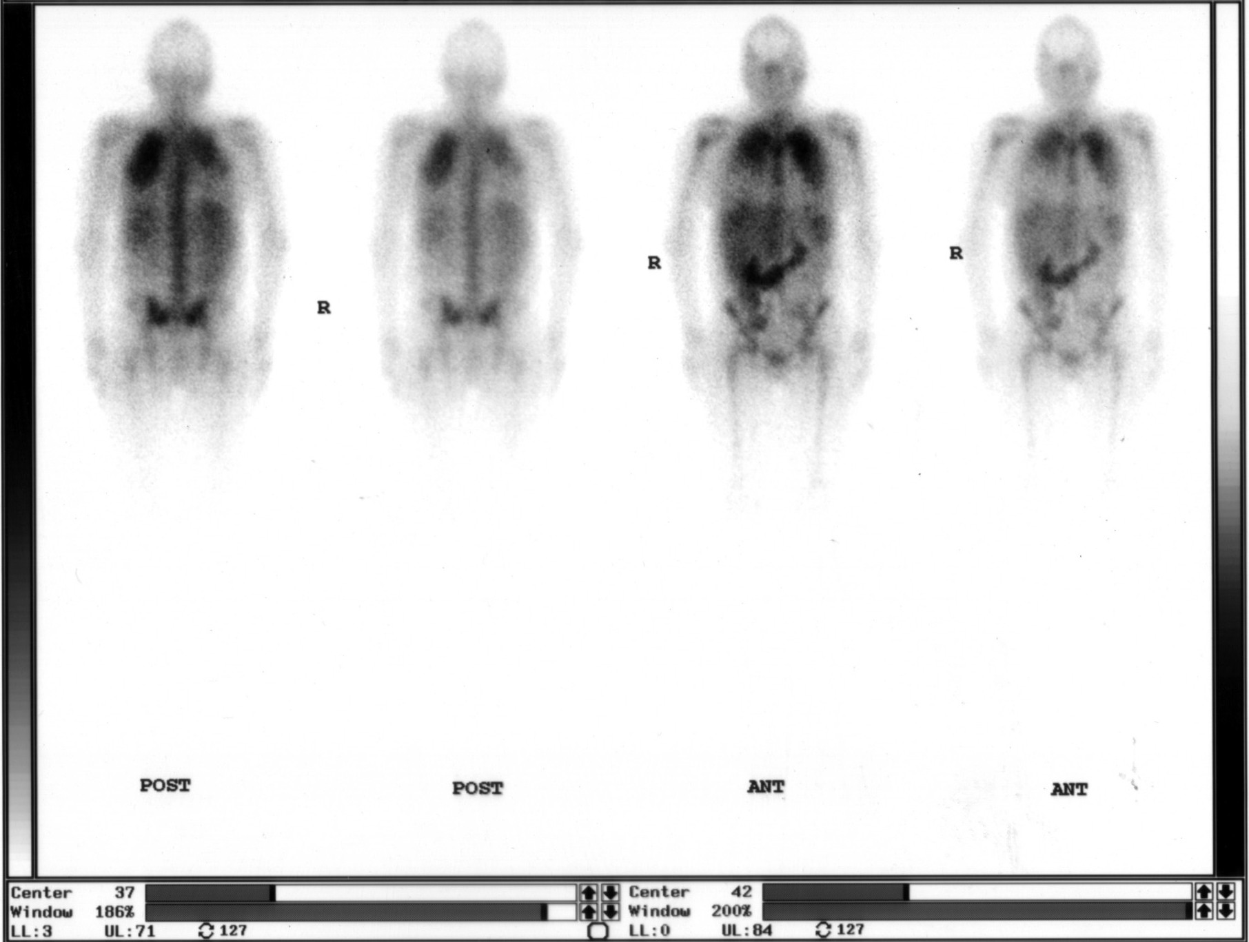 Ga-67 Citrate Whole-Body Scan: Agent and Timing
Abnormal Bilateral Lung Uptake on Gallium Scan
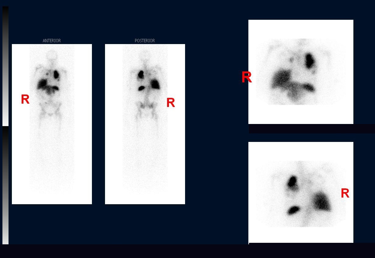 In-111 Oxine WBC Scan: Agent and Timing
Abnormal Lung Uptake on In-111 WBC Scan
All 130 nuclear medicine images by category →
Practise image interpretation in the app
All 130 images as an interactive quiz, alongside 500 NMTCB and ARRT(N) questions with worked explanations, case studies and timed mock exams. 25 questions are free, no card and no account.