Melanoma Lymphoscintigraphy: The Immediate Image
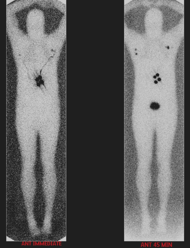
Provided clinical image
Question
This melanoma lymphoscintigraphy shows two images side by side: an immediate image taken right after the peritumoral injection and an anterior 45-minute delayed image. What is the PRIMARY purpose of obtaining the IMMEDIATE image?
- ATo measure leftover activity in the syringe and calculate the exact dose administered to the patient
- BTo image the blood flow phase of the radiotracer as it enters the lymphatic system from the injection site
- CTo check that the patient is properly positioned in the field of view before the delayed images are acquired
- DTo confirm the injection was successful and mark the primary tumor site so it can be distinguished from sentinel nodes on delayed images
Show the answer and explanation
D. To confirm the injection was successful and mark the primary tumor site so it can be distinguished from sentinel nodes on delayed images
The immediate image in lymphoscintigraphy serves as documentation that the tracer was successfully injected at the correct site (around the melanoma or biopsy scar). It establishes the baseline injection site location so that when sentinel nodes appear on delayed images, the technologist and surgeon can clearly distinguish nodes from the injection site. It also helps the surgeon understand the anatomical relationship between the primary tumor and the draining sentinel nodes.
More lymphoscintigraphy images
Melanoma Lymphoscintigraphy: Multiple Draining Basins
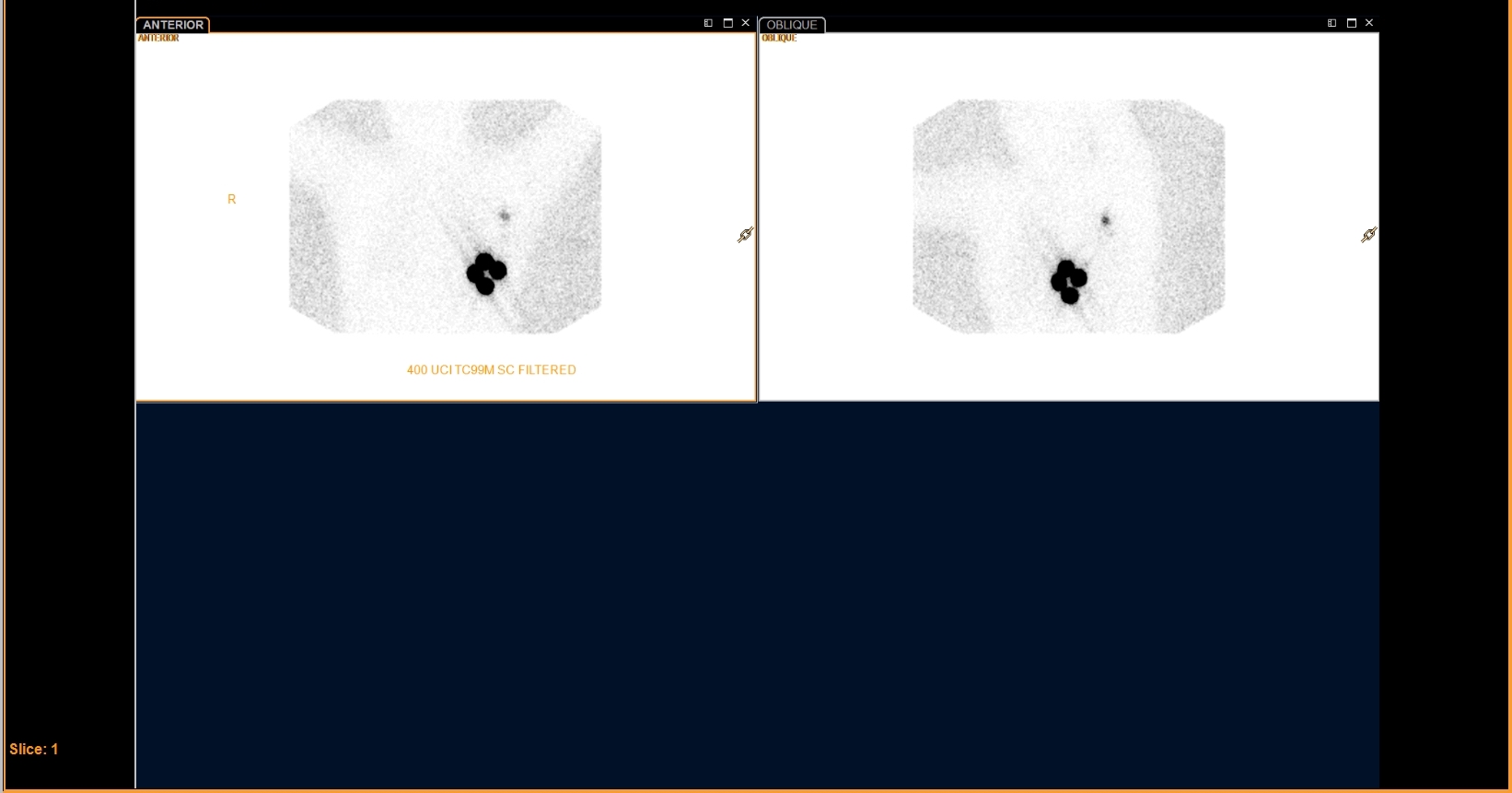 Filtered Tc-99m Sulfur Colloid for Breast Lymphoscintigraphy
Breast Lymphoscintigraphy: Why Oblique Views
All 130 nuclear medicine images by category →
Practise image interpretation in the app
All 130 images as an interactive quiz, alongside 500 NMTCB and ARRT(N) questions with worked explanations, case studies and timed mock exams. 25 questions are free, no card and no account.