Breast Lymphoscintigraphy: Why Oblique Views
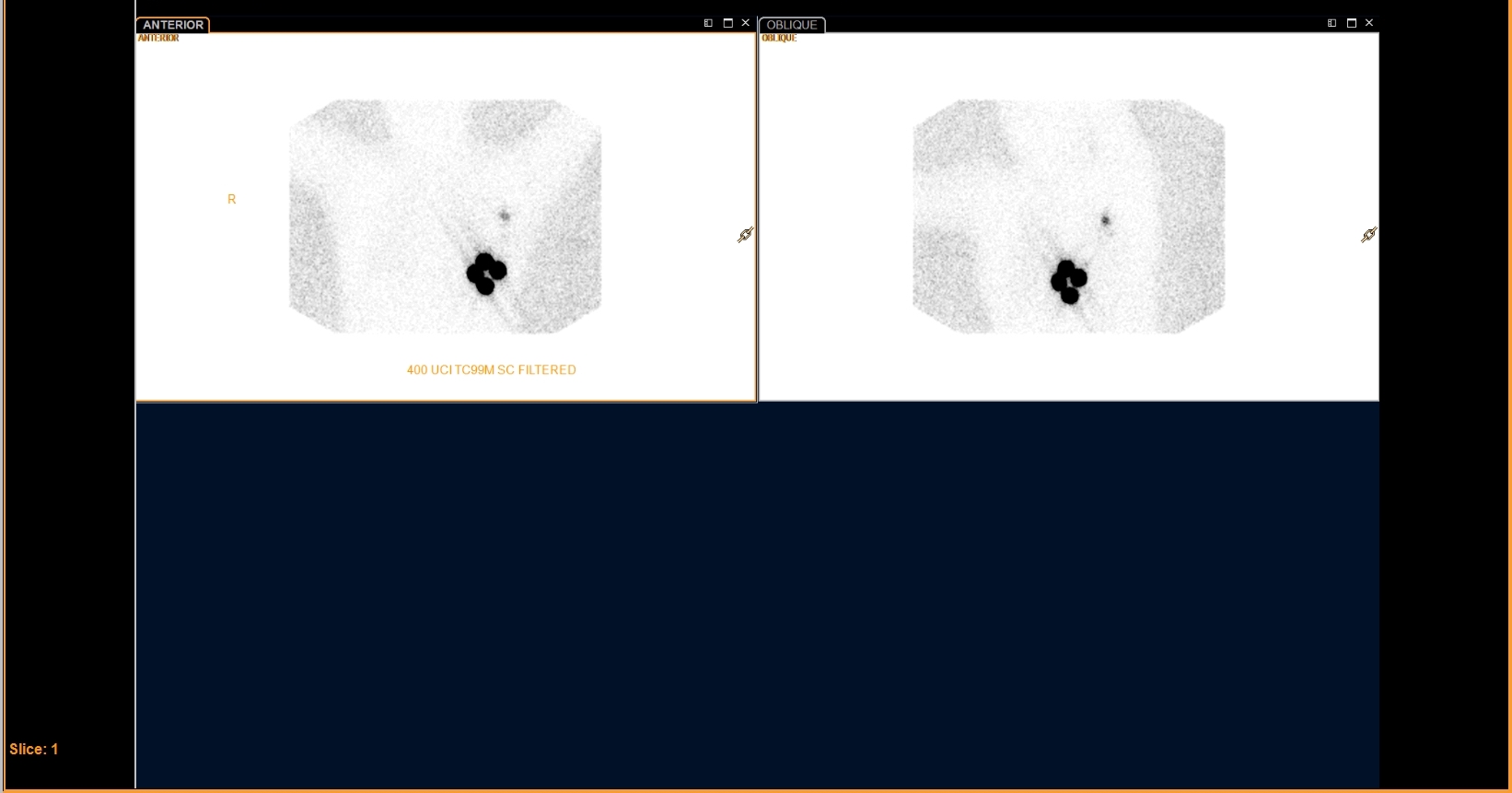
Provided clinical image
Question
This breast lymphoscintigraphy includes both anterior and oblique views of the axilla. What is the PRIMARY reason for acquiring images in more than one projection?
- ATo double the imaging time per view so that enough counts are collected in the sentinel node for accurate quantification of radiotracer uptake
- BRadiation safety protocols require two projections to confirm that tracer has cleared sufficiently from the injection site before the patient is released
- CMultiple views help localize the sentinel lymph node in three dimensions and separate it from the injection site, giving the surgeon accurate depth and position information
- DComparing both projections allows the technologist to identify bilateral axillary sentinel nodes by detecting asymmetry between the two sides
Show the answer and explanation
C. Multiple views help localize the sentinel lymph node in three dimensions and separate it from the injection site, giving the surgeon accurate depth and position information
Acquiring anterior and oblique (or lateral) views during lymphoscintigraphy serves two purposes: (1) it allows the sentinel node to be separated from the bright injection site, which can otherwise overlap the node on a single anterior view; and (2) it gives the surgeon three-dimensional localization of the sentinel node before the procedure. The technologist typically marks the skin over the sentinel node using a transmission marker or a radioactive point source, and the oblique view confirms the exact position. This information guides the surgeon's gamma probe intraoperatively.
More lymphoscintigraphy images
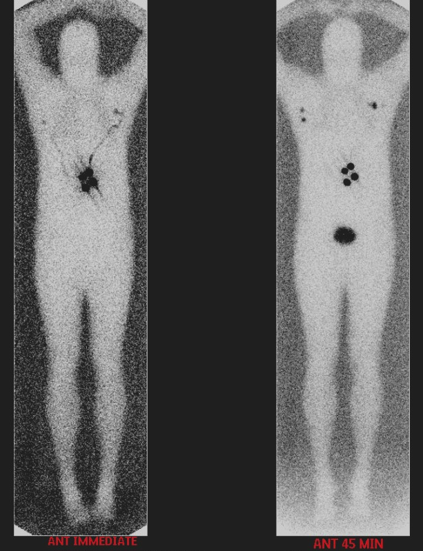 Melanoma Lymphoscintigraphy: The Immediate Image
Melanoma Lymphoscintigraphy: Multiple Draining Basins
Filtered Tc-99m Sulfur Colloid for Breast Lymphoscintigraphy
All 130 nuclear medicine images by category →
Practise image interpretation in the app
All 130 images as an interactive quiz, alongside 500 NMTCB and ARRT(N) questions with worked explanations, case studies and timed mock exams. 25 questions are free, no card and no account.