Filtered Tc-99m Sulfur Colloid for Breast Lymphoscintigraphy
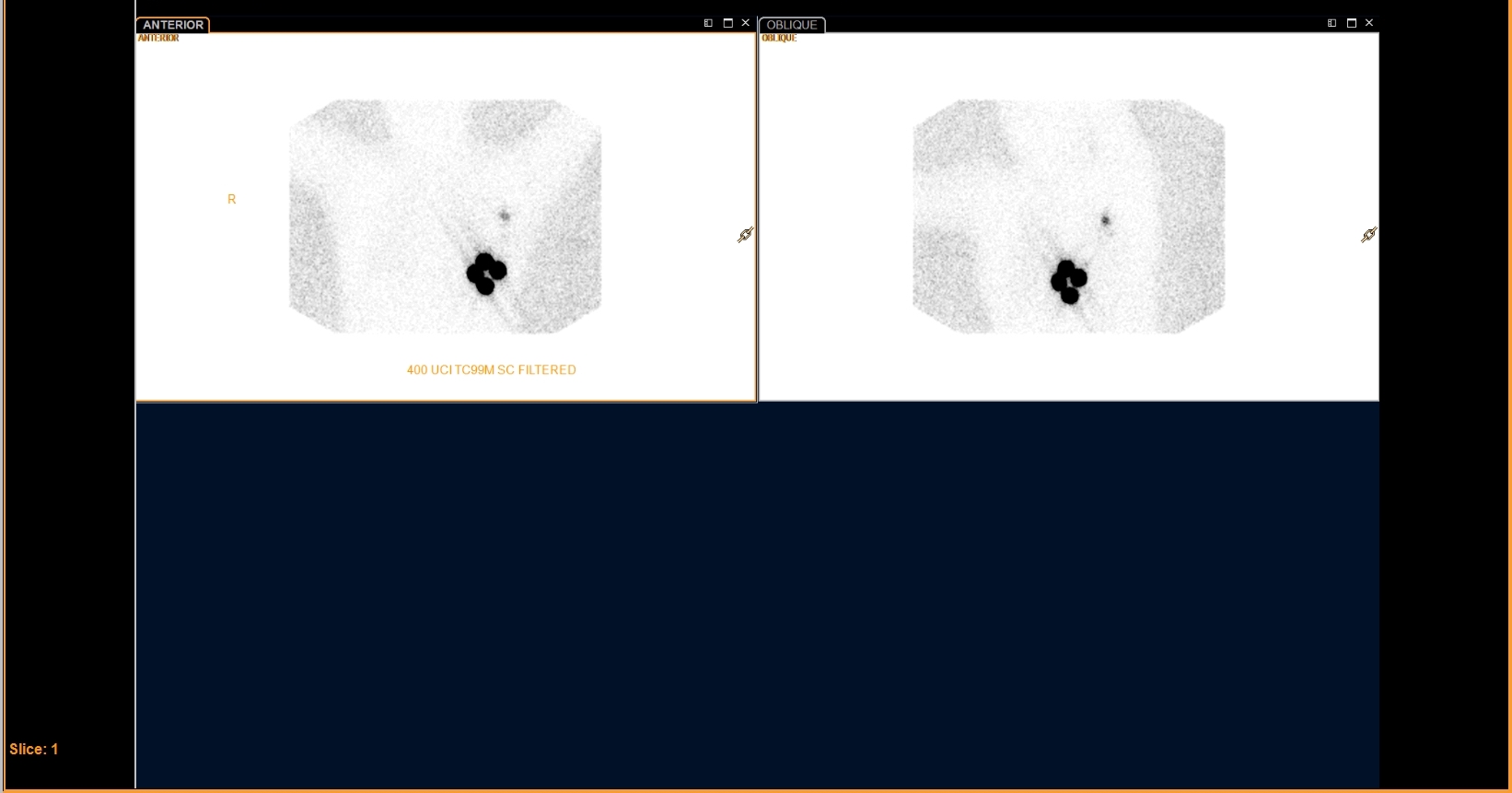
Provided clinical image
Question
The image label reads '400 UCI TC99M SC FILTERED.' Why is FILTERED Tc-99m sulfur colloid used for breast lymphoscintigraphy rather than unfiltered sulfur colloid?
- AFiltering removes free pertechnetate that would otherwise migrate too quickly through the lymphatics and produce excessive background activity throughout the chest
- BFiltered sulfur colloid has a longer shelf life and remains stable for up to 8 hours after preparation, making it more practical for procedure scheduling
- CFiltering increases particle size so the tracer stays at the injection site longer, giving the surgeon more time to locate the sentinel node intraoperatively
- DFiltering produces smaller particles (0.1–0.2 micron) that travel more efficiently through the lymphatic channels to reach the sentinel lymph node
Show the answer and explanation
D. Filtering produces smaller particles (0.1–0.2 micron) that travel more efficiently through the lymphatic channels to reach the sentinel lymph node
Unfiltered Tc-99m sulfur colloid has a wide range of particle sizes (up to 1 micron), and larger particles tend to remain at the injection site rather than migrating through lymphatics. Filtering through a 0.1–0.22 micron membrane produces a uniform smaller-particle preparation that moves more readily through the lymphatic system to the sentinel node, improving image quality and sentinel node identification. This is why filtered sulfur colloid is the standard radiopharmaceutical for lymphoscintigraphy in the United States.
More lymphoscintigraphy images
Breast Lymphoscintigraphy: Why Oblique Views
 Melanoma Lymphoscintigraphy: The Immediate Image
Melanoma Lymphoscintigraphy: Multiple Draining Basins
All 130 nuclear medicine images by category →
Practise image interpretation in the app
All 130 images as an interactive quiz, alongside 500 NMTCB and ARRT(N) questions with worked explanations, case studies and timed mock exams. 25 questions are free, no card and no account.