Melanoma Lymphoscintigraphy: Multiple Draining Basins
Provided clinical image
Question
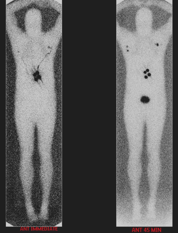
On the delayed images from this truncal melanoma lymphoscintigraphy, sentinel lymph nodes appear in more than one lymph node basin. As the nuclear medicine technologist, what is the significance of reporting ALL identified draining basins to the surgeon?
- AMultiple draining basins indicate the lymphoscintigraphy failed and the injection should be repeated at a different location around the melanoma site
- BEach draining basin identified on the scan must be surgically explored, because melanoma can spread to any sentinel node in any basin that shows tracer drainage from the tumor
- CMultiple basins indicate the melanoma has already spread to distant lymph nodes, automatically changing the patient's staging to Stage IV metastatic disease
- DMultiple basins on the scan mean the patient received too large a radiotracer dose, causing nonspecific uptake in lymph nodes unrelated to the primary melanoma drainage
Show the answer and explanation
B. Each draining basin identified on the scan must be surgically explored, because melanoma can spread to any sentinel node in any basin that shows tracer drainage from the tumor
Truncal melanomas (located on the torso) can drain unpredictably to multiple lymph node basins — for example, both axillae, or one axilla and the ipsilateral groin. Unlike extremity melanomas that typically drain in one direction, truncal melanomas may drain to any basin on either side of the body. The lymphoscintigraphy map tells the surgeon exactly which basins to explore intraoperatively with a gamma probe. If the technologist fails to report all identified draining basins, the surgeon may miss a sentinel node containing micrometastatic disease.
More lymphoscintigraphy images
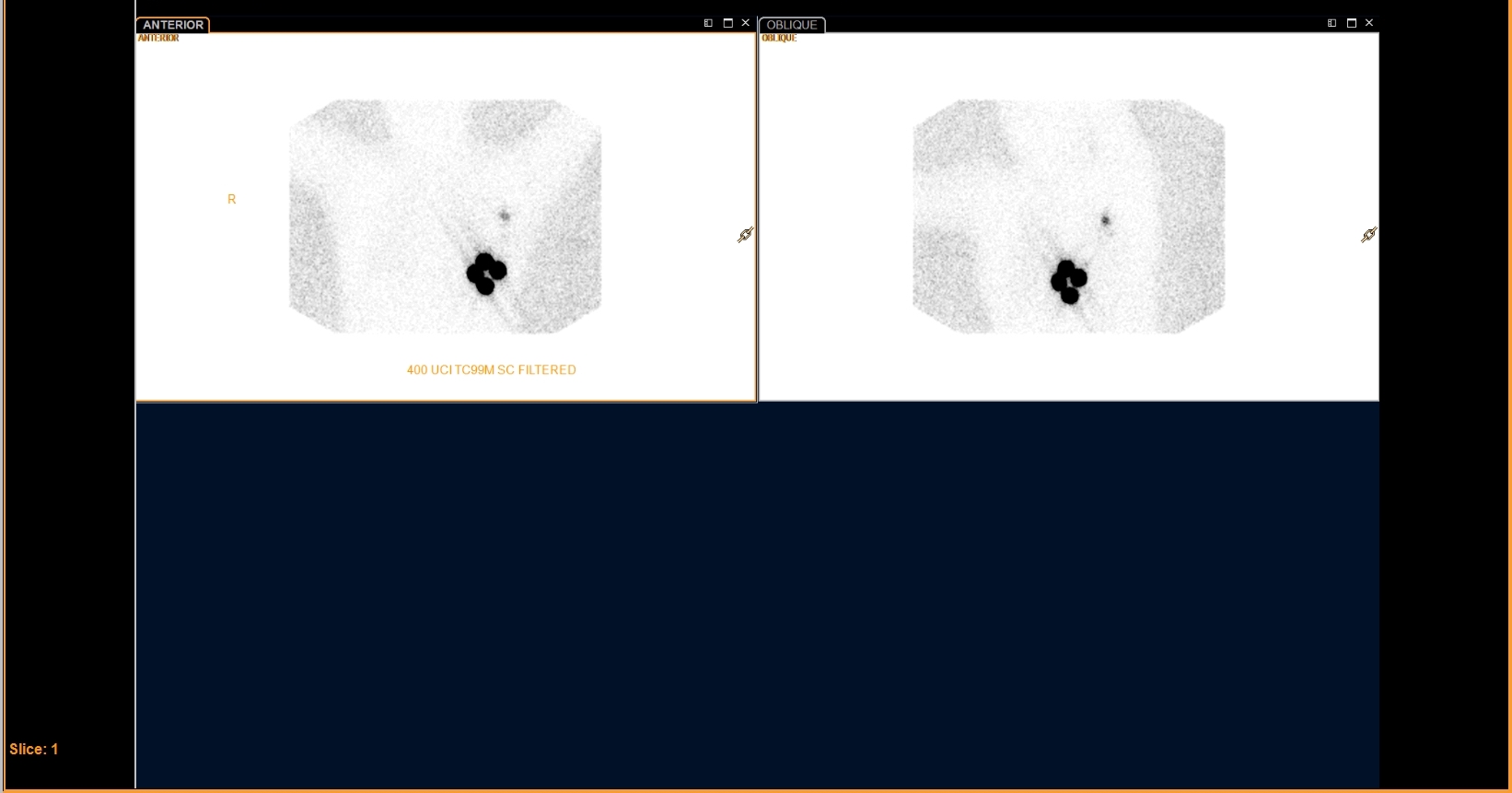 Filtered Tc-99m Sulfur Colloid for Breast Lymphoscintigraphy
Breast Lymphoscintigraphy: Why Oblique Views
Melanoma Lymphoscintigraphy: The Immediate Image
All 130 nuclear medicine images by category →
Practise image interpretation in the app
All 130 images as an interactive quiz, alongside 500 NMTCB and ARRT(N) questions with worked explanations, case studies and timed mock exams. 25 questions are free, no card and no account.